
Human VAP-1/AOC3 Antibody
R&D Systems, part of Bio-Techne | Catalog # MAB3957

Scientific images may be resized, cropped, or adjusted for brightness or contrast for presentation purposes. These updates are limited to presentation and do not affect the underlying scientific interpretation of the data. Where available, additional source data may be provided upon request.
Conjugate
Catalog #
Key Product Details
Species Reactivity
Validated:
Human
Cited:
Human
Applications
Validated:
Western Blot
Cited:
Immunohistochemistry, Immunohistochemistry-Paraffin, Western Blot
Label
Unconjugated
Antibody Source
Monoclonal Mouse IgG2B Clone # 393112
Product Specifications
Immunogen
S. frugiperda insect ovarian cell line Sf 21-derived recombinant human VAP‑1/AOC3
Gly27-Asn763
Accession # Q16853
Gly27-Asn763
Accession # Q16853
Specificity
Detects human VAP‑1/AOC3 in direct ELISAs and Western blots. In Western blots, 50% cross-reactivity with recombinant mouseVAP‑1/AOC3 is observed.
Clonality
Monoclonal
Host
Mouse
Isotype
IgG2B
Scientific Data Images for Human VAP-1/AOC3 Antibody
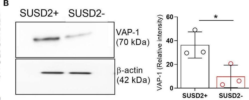
Detection of VAP-1/AOC3 by Western Blot
Biophysical and functional characterization of endometrial pericytes and EnSCs. (A) Immunohistochemistry showing VAP-1 expression in luteal-phase human endometrium, hematoxylin was used to stain the nuclei. Staining indicates VAP-1 reactivity in the vasculature and spiral arteries, arrow pointing to spiral artery. Scale bar: 200 μm. (B) VAP-1 expression was determined at protein level in freshly isolated SUSD2+ pericytes and SUSD2− EnSCs from three independent endometrial biopsies by Western blot analysis and normalized to beta-actin and relative intensity determined by densitometry; Student's t-test; *P < 0.05. (C) Expression of ELN (left panel), CNN1 (middle panel) and MYH11 (right panel) transcripts was determined by RT-qPCR in freshly isolated SUSD2+ pericytes and SUSD2− EnSCs from four independent endometrial biopsies; Student's t-test; *P < 0.05; **P < 0.01. (D) Representative graphs showing proliferation and migration SUSD2+ pericytes and SUSD2− EnSCs monitored in real-time using the xCELLigence system for the indicated time-points. (E) freshly isolated SUSD2+ pericytes and SUSD2− EnSCs were embedded into collagen at a density of 1 × 10⁶ cells per gel. Single cell contraction force was measured using the depth-sensing nanoindentation system. Data represent mean ± SEM of four biological repeat experiments. Student's t-test; **P < 0.01; ****P < 0.0001. Image collected and cropped by CiteAb from the following open publication (https://pubmed.ncbi.nlm.nih.gov/33537312), licensed under a CC-BY license. Not internally tested by R&D Systems.Applications for Human VAP-1/AOC3 Antibody
Application
Recommended Usage
Western Blot
1 µg/mL
Sample: Recombinant Human VAP‑1/AOC3 (Catalog # 3957-AO) under reducing conditions only
Sample: Recombinant Human VAP‑1/AOC3 (Catalog # 3957-AO) under reducing conditions only
Reviewed Applications
Read 1 review rated 3 using MAB3957 in the following applications:
Formulation, Preparation, and Storage
Purification
Protein A or G purified from hybridoma culture supernatant
Reconstitution
Reconstitute at 0.5 mg/mL in sterile PBS. For liquid material, refer to CoA for concentration.
Formulation
Lyophilized from a 0.2 μm filtered solution in PBS with Trehalose. *Small pack size (SP) is supplied either lyophilized or as a 0.2 µm filtered solution in PBS.
Shipping
Lyophilized product is shipped at ambient temperature. Liquid small pack size (-SP) is shipped with polar packs. Upon receipt, store immediately at the temperature recommended below.
Stability & Storage
Use a manual defrost freezer and avoid repeated freeze-thaw cycles.
- 12 months from date of receipt, -20 to -70 °C as supplied.
- 1 month, 2 to 8 °C under sterile conditions after reconstitution.
- 6 months, -20 to -70 °C under sterile conditions after reconstitution.
Background: VAP-1/AOC3
Vascular adhesion protein-1 (VAP-1), also called AOC3 (amine oxidase copper-containing 3) or SSAO (semicarbazide-sensitive amine oxidase), is a copper amine oxidase with a topaquinone cofactor. VAP-1 is a Type II integral membrane protein, but a soluble form of the enzyme is present in human serum, and its level increases in diabetes and some inflammatory liver diseases (1, 2). Human and mouse VAP-1 share 83% amino acid sequence identity. VAP-1 catalyzes the oxidative deamination of small primary amines such as methylamine, benzylamine, and aminoacetone in a reaction that produces an aldehyde, ammonia, and H2O2 (3). The enzyme is sensitive to inhibition by semicarbazide. VAP-1 expression is highest in the endothelium of lung, heart, and intestine, but low in tissues such as brain, spleen, kidney, and liver (4). VAP-1 vascular expression is regulated at sites of inflammation through its release from intracellular granules in which the protein is stored (5). The adhesive function of VAP-1 has been demonstrated in studies showing that the protein is important for the adherence of certain lymphocyte subtypes to inflamed endothelial tissues (6). VAP-1 mediated adhesion is involved in the process of leukocyte extravasation, an important feature of inflammatory responses. The role of VAP-1 amine oxidase activity in this process is not fully defined, but it appears to be carbohydrate-dependent (7). VAP-1 is considered to be a therapeutic target for diabetes, oxidative stress, and inflammatory diseases (8).
References
- Kurkijärvi, R. et al. (1998) J. Immunol. 161:1549.
- Gearing, A.J.H. and W. Newman (1993) Immunol. Today 14:506.
- Lizcano, J.M. et al. (1998) Biochem. J. 331:69.
- Smith, D.J. et al. (1998) J. Exp. Med. 188:17.
- Jaakkala K. et al. (2000) Am. J. Pathol. 157:463.
- Salmi, M. and J. Jalkanen (2001) Trends Immunol. 22:211.
- Salmi, M. and J. Jalkanen (1996) J. Exp. Med. 183:569.
- Dunkel, P. et al. (2008) Curr. Med. Chem. 15:1827.
Long Name
Vascular Adhesion Protein-1
Alternate Names
AOC3, HPAO, SSAO, VAP1
Gene Symbol
AOC3
UniProt
Additional VAP-1/AOC3 Products
Product Documents for Human VAP-1/AOC3 Antibody
Product Specific Notices for Human VAP-1/AOC3 Antibody
For research use only
Loading...
Loading...
Loading...
Loading...
Loading...