
Human Phospho-FGFR1-4 (Y653/Y654) Antibody
R&D Systems, part of Bio-Techne | Catalog # AF3285


Scientific images may be resized, cropped, or adjusted for brightness or contrast for presentation purposes. These updates are limited to presentation and do not affect the underlying scientific interpretation of the data. Where available, additional source data may be provided upon request.
Key Product Details
Validated by
Biological Validation
Species Reactivity
Validated:
Human
Cited:
Human, Mouse, Xenograft
Applications
Validated:
Western Blot, Immunocytochemistry
Cited:
Western Blot, Immunocytochemistry, Immunocytochemistry/ Immunofluorescence, Immunoprecipitation
Label
Unconjugated
Antibody Source
Polyclonal Rabbit IgG
Product Specifications
Immunogen
Phosphopeptide containing human FGF R1 Y653/Y654 sites
Specificity
Detects human FGF R when dually phosphorylated at Y653/Y654 in Western blots.
Clonality
Polyclonal
Host
Rabbit
Isotype
IgG
Scientific Data Images for Human Phospho-FGFR1-4 (Y653/Y654) Antibody
Detection of Human Phospho- FGF R1‑4 (Y653/Y654) by Western Blot.
Western blot shows lysates of KATO-III human gastric carcinoma cell line untreated (-) or treated (+) with 100 ng/mL Recom-binant Human FGF acidic (Catalog # 232-FA) for 15 minutes. PVDF membrane was probed with 0.5 µg/mL of Rabbit Anti-Human Phospho-FGF R1-4 (Y653/Y654) Antigen Affinity-purified Polyclonal Antibody (Catalog # AF3285), followed by HRP-conjugated Anti-Rabbit IgG Secondary Antibody (Catalog # HAF008). A specific band was detected for Phospho-FGF R1-4 (Y653/Y654) at approximately 120 to 145 kDa (as indicated). This experiment was conducted under reducing conditions and using Immunoblot Buffer Group 1.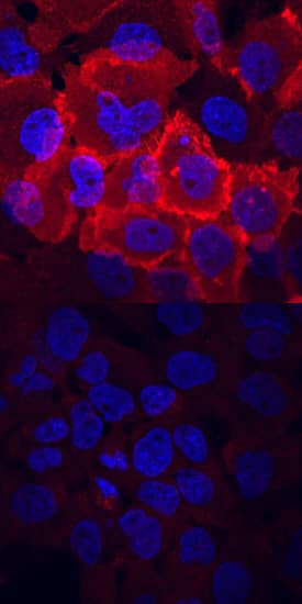
Phospho-FGF R1-4 (Y653/Y654) in A431 Human Cell Line.
FGF R1-4 phosphorylated at Y653/Y654 was detected in immersion fixed A431 human epithelial carcinoma cell line untreated (lower panel) or treated (upper panel) with pervanadate using Rabbit Anti-Human Phospho-FGF R1-4 (Y653/Y654) Antigen Affinity-purified Polyclonal Antibody (Catalog # AF3285) at 10 µg/mL for 3 hours at room temperature. Cells were stained using the NorthernLights™ 557-conjugated Anti-Rabbit IgG Secondary Antibody (red; Catalog # NL004) and counterstained with DAPI (blue). View our protocol for Fluorescent ICC Staining of Cells on Coverslips.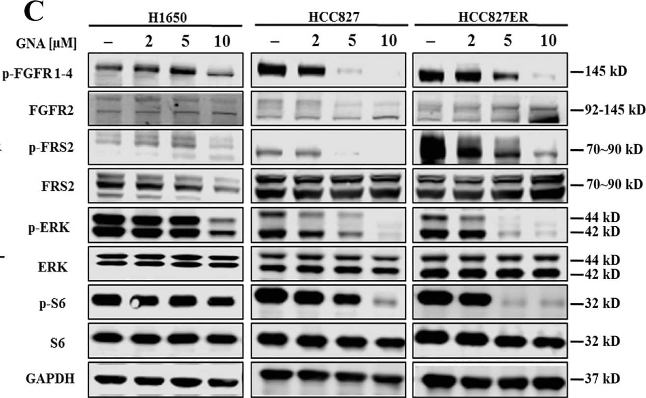
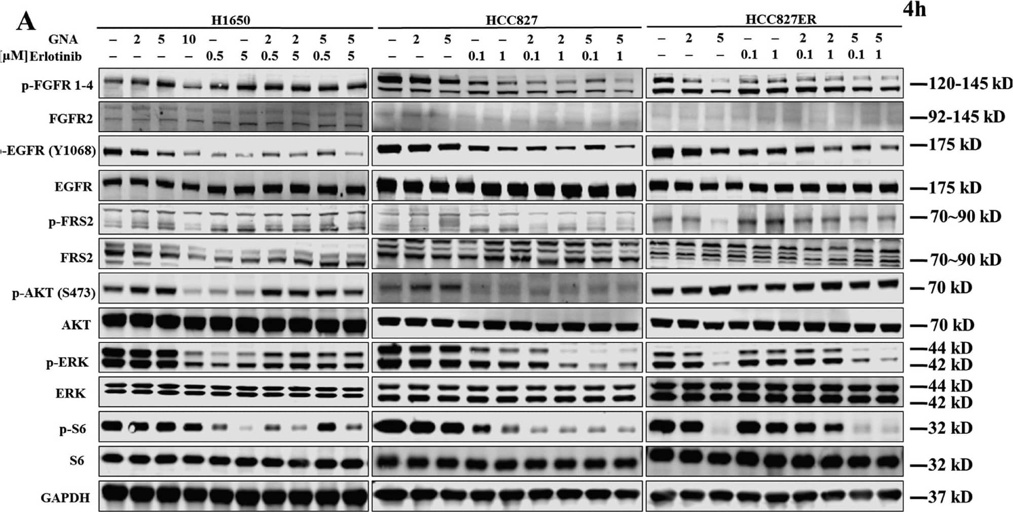
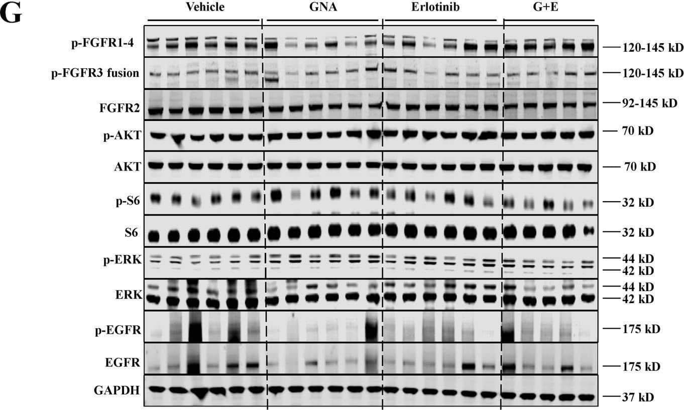
Detection of Phospho-FGFR1-4 (Y653/Y654) by Western Blot
GNA inhibits cell proliferation in both NSCLC and erlotinib-resistant NSCLC by targeting FGFR and the downstream signaling pathways.a Cell viability was assessed using the CellTiter-Glo assay. After a 72 h treatment by GNA, CellTiter-Glo reagent was added directly to each well for a 10-min incubation. The plate was then read on a FlexStation 3 microplate luminometer. A dose−response curve was plotted, and the IC50 was calculated using GraphPad Prism software. Cell viability was assessed using MTT assay. After a 72 h treatment by GNA, 10 μl of 5 mg/ml 3-(4,5-dimethylthiazol-2-yl)-2,5-diphenyltetrazolium bromide (MTT) solution was added to each well and incubated for 4 h at 37 °C, then the culture medium was removed and 100 μl of DMSO was added to each well. The absorbance was measured at 570 nm on a microplate reader. A dose−response curve was plotted, and the IC50 was calculated using GraphPad Prism software. b Caspase-3/7 activity induction was evaluated after 6 h of treatment with GNA at 10, 3.33 and 1.11 μM using the Caspase-Glo 3/7 kit from Promega in the HCC827, H1650, and HCC827ER cells, respectively. The bars in the graphs represent the mean fold induction relative to the DMSO control. c H1650 cells, HCC827 cells, and HCC827ER cells were treated with GNA at various dosages for 4 h and were then probed with specified antibodies. FGFR and the downstream proteins were analyzed by western blot. d The histograms of quantitative analysis about FGFR and the downstream proteins were analyzed by western blot Image collected and cropped by CiteAb from the following open publication (https://pubmed.ncbi.nlm.nih.gov/29449529), licensed under a CC-BY license. Not internally tested by R&D Systems.Applications for Human Phospho-FGFR1-4 (Y653/Y654) Antibody
Application
Recommended Usage
Immunocytochemistry
5-15 µg/mL
Sample: Immersion fixed A431 human epithelial carcinoma cell line treated with pervanadate
Sample: Immersion fixed A431 human epithelial carcinoma cell line treated with pervanadate
Western Blot
0.5 µg/mL
Sample: KATO-III human gastric carcinoma cell line treated with Recombinant Human FGF acidic (Catalog # 232-FA)
Sample: KATO-III human gastric carcinoma cell line treated with Recombinant Human FGF acidic (Catalog # 232-FA)
Formulation, Preparation, and Storage
Purification
Antigen Affinity-purified
Reconstitution
Reconstitute at 0.2 mg/mL in sterile PBS. For liquid material, refer to CoA for concentration.
Formulation
Lyophilized from a 0.2 μm filtered solution in PBS with Trehalose. *Small pack size (SP) is supplied either lyophilized or as a 0.2 µm filtered solution in PBS.
Shipping
Lyophilized product is shipped at ambient temperature. Liquid small pack size (-SP) is shipped with polar packs. Upon receipt, store immediately at the temperature recommended below.
Stability & Storage
Use a manual defrost freezer and avoid repeated freeze-thaw cycles.
- 12 months from date of receipt, -20 to -70 °C as supplied.
- 1 month, 2 to 8 °C under sterile conditions after reconstitution.
- 6 months, -20 to -70 °C under sterile conditions after reconstitution.
Background: FGFR1-4
Long Name
Fibroblast Growth Factor Receptors 1-4
Alternate Names
BFGFR;bFGF-R-1;CD331;CEK;ECCL;FGFBR;FGFR-1;Fibroblast growth factor receptor;Fibroblast growth factor receptor 1;FLG;FLT2;FLT-2;HBGFR;HH2;HRTFDS;KAL2;N-SAM;OGD
Additional FGFR1-4 Products
Product Documents for Human Phospho-FGFR1-4 (Y653/Y654) Antibody
Product Specific Notices for Human Phospho-FGFR1-4 (Y653/Y654) Antibody
For research use only
Loading...
Loading...
Loading...
Loading...
Loading...