
Human PDGF-D Antibody
R&D Systems, part of Bio-Techne | Catalog # AF1159

Scientific images may be resized, cropped, or adjusted for brightness or contrast for presentation purposes. These updates are limited to presentation and do not affect the underlying scientific interpretation of the data. Where available, additional source data may be provided upon request.
Conjugate
Catalog #
Key Product Details
Species Reactivity
Validated:
Human
Cited:
Human
Applications
Validated:
Western Blot
Cited:
Immunohistochemistry, Immunohistochemistry-Frozen, Western Blot, Neutralization, Immunoprecipitation
Label
Unconjugated
Antibody Source
Polyclonal Goat IgG
Product Specifications
Immunogen
E. coli-derived recombinant human PDGF-D
Asp253-Arg370
Accession # Q9GZP0
Asp253-Arg370
Accession # Q9GZP0
Specificity
Detects human PDGF-D in direct ELISAs and Western blots. In direct ELISAs and Western blots, less than 1% cross-reactivity with recombinant human (rh) PDGF-AA, rhPDGF-AB and rhPDGF-BB is observed.
Clonality
Polyclonal
Host
Goat
Isotype
IgG
Scientific Data Images for Human PDGF-D Antibody
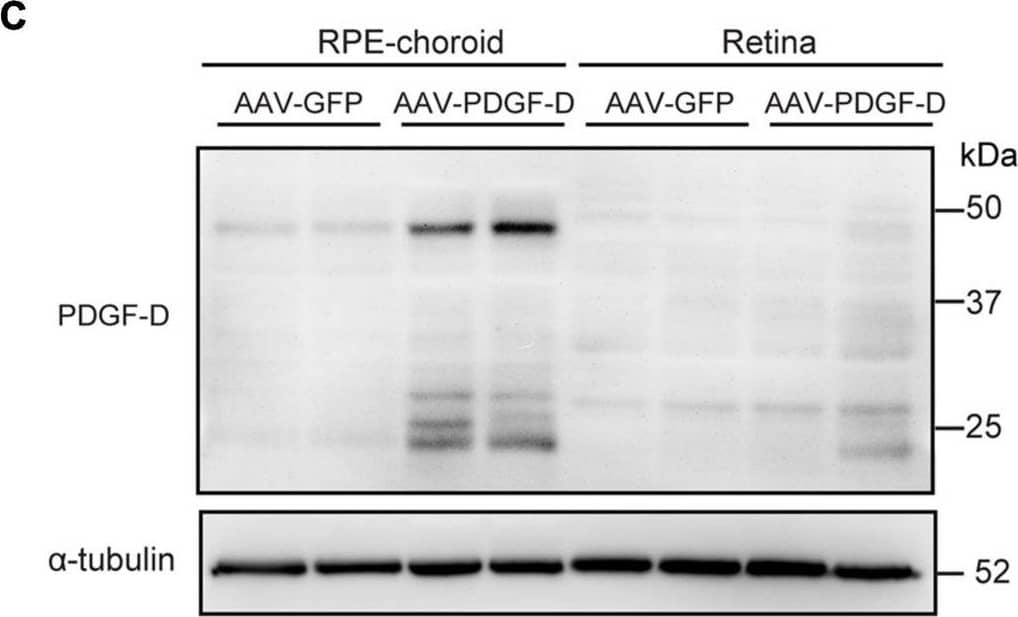
Detection of Mouse PDGF-D/SCDGFB by Western Blot
RPE-specific overexpression of PDGF-D in vivo.(A) Schematic diagram illustrating the AAV vector carrying GFP (AAV-GFP) or human PDGF-D (AAV-PDGF-D) gene and RPE specific VDM2 promoter. (B) Real-time PCR results of relative mRNA expression of PDGF-D in retinae or RPE-choroid complex from mice injected with AAV-GFP or AAV-PDGF- D for 4 weeks. (C) Representative immunoblotting showing PDGF-D expression in mouse retinae or RPE-choroid complex. (D) Quantifications of PDGF-D protein levels in (C). (E) Immunofluorescence images highlighting RPE-specific expression of GFP or PDGF-D in mouse RPE-choroid complex with 4 weeks AAV-GFP or AAV-PDGF-D injection, respectively. Scale bar: 50 μm. n = 5, ****p < 0.0001, ns: not significant. Image collected and cropped by CiteAb from the following open publication (https://pubmed.ncbi.nlm.nih.gov/34150781), licensed under a CC-BY license. Not internally tested by R&D Systems.Applications for Human PDGF-D Antibody
Application
Recommended Usage
Western Blot
0.1 µg/mL
Sample: Recombinant Human PDGF-D
Sample: Recombinant Human PDGF-D
Formulation, Preparation, and Storage
Purification
Antigen Affinity-purified
Reconstitution
Reconstitute at 0.2 mg/mL in sterile PBS. For liquid material, refer to CoA for concentration.
Formulation
Lyophilized from a 0.2 μm filtered solution in PBS with Trehalose. See Certificate of Analysis for details.
*Small pack size (-SP) is supplied either lyophilized or as a 0.2 µm filtered solution in PBS.
*Small pack size (-SP) is supplied either lyophilized or as a 0.2 µm filtered solution in PBS.
Shipping
Lyophilized product is shipped at ambient temperature. Liquid small pack size (-SP) is shipped with polar packs. Upon receipt, store immediately at the temperature recommended below.
Stability & Storage
Use a manual defrost freezer and avoid repeated freeze-thaw cycles.
- 12 months from date of receipt, -20 to -70 °C as supplied.
- 1 month, 2 to 8 °C under sterile conditions after reconstitution.
- 6 months, -20 to -70 °C under sterile conditions after reconstitution.
Background: PDGF-D
Long Name
Platelet-derived Growth Factor D
Alternate Names
PDGFD
Gene Symbol
PDGFD
UniProt
Additional PDGF-D Products
Product Documents for Human PDGF-D Antibody
Product Specific Notices for Human PDGF-D Antibody
For research use only
Loading...
Loading...
Loading...
Loading...
Loading...