
Human Nidogen-1/Entactin Antibody
R&D Systems, part of Bio-Techne | Catalog # AF2570



Scientific images may be resized, cropped, or adjusted for brightness or contrast for presentation purposes. These updates are limited to presentation and do not affect the underlying scientific interpretation of the data. Where available, additional source data may be provided upon request.
Conjugate
Catalog #
Key Product Details
Species Reactivity
Validated:
Human
Cited:
Human, Mouse, Rabbit
Applications
Validated:
Immunohistochemistry, Western Blot
Cited:
Immunohistochemistry, Immunohistochemistry-Frozen, Western Blot, Immunocytochemistry
Label
Unconjugated
Antibody Source
Polyclonal Goat IgG
Product Specifications
Immunogen
Mouse myeloma cell line NS0-derived recombinant human Nidogen‑1/Entactin
Leu29-Lys1114 (Gln1113Arg)
Accession # AAH45606
Leu29-Lys1114 (Gln1113Arg)
Accession # AAH45606
Specificity
Detects human Nidogen‑1/Entactin in direct ELISAs and Western blots.
Clonality
Polyclonal
Host
Goat
Isotype
IgG
Scientific Data Images for Human Nidogen-1/Entactin Antibody
Nidogen-1/Entactin in Human Heart.
Nidogen-1/Entactin was detected in immersion fixed paraffin-embedded sections of human heart using Goat Anti-Human Nidogen-1/Entactin Antigen Affinity-purified Polyclonal Antibody (Catalog # AF2570) at 15 µg/mL overnight at 4 °C. Tissue was stained using the Anti-Goat HRP-DAB Cell & Tissue Staining Kit (brown; Catalog # CTS008) and counterstained with hematoxylin (blue). Specific labeling was localized to the sarcolemma of cardiomyocytes. View our protocol for Chromogenic IHC Staining of Paraffin-embedded Tissue Sections.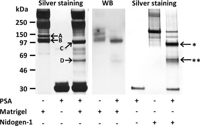
Detection of Human Nidogen-1/Entactin by Western Blot
Degradation of nidogen-1 by PSA.PSA cleaved both mouse nidogen-1 in Matrigel and human recombinant nidogen-1. Mass spectrometry analysis identified nidogen-1 in the silver stained gel bands (arrows, left panel). Nidogen-1 bands of 140 kDa (A) and 110 kDa (B) disappeared and fragments of 90 kDa (C) and 55 kDa (D) appeared after 48 h incubation of diluted Matrigel with PSA at 37°C. Nidogen-1 cleavage by PSA in Matrigel was visualized by Western blotting with anti-nidogen-1 polyclonal antibody (middle panel). PSA (1 µM) cleaved recombinant human nidogen-1 (0.5 µM) into two fragments 85 kDa (arrow with *) and 55 kDa (arrow with **) during 20 h incubation at 37°C (right panel). Image collected and cropped by CiteAb from the following publication (https://dx.plos.org/10.1371/journal.pone.0107819), licensed under a CC-BY license. Not internally tested by R&D Systems.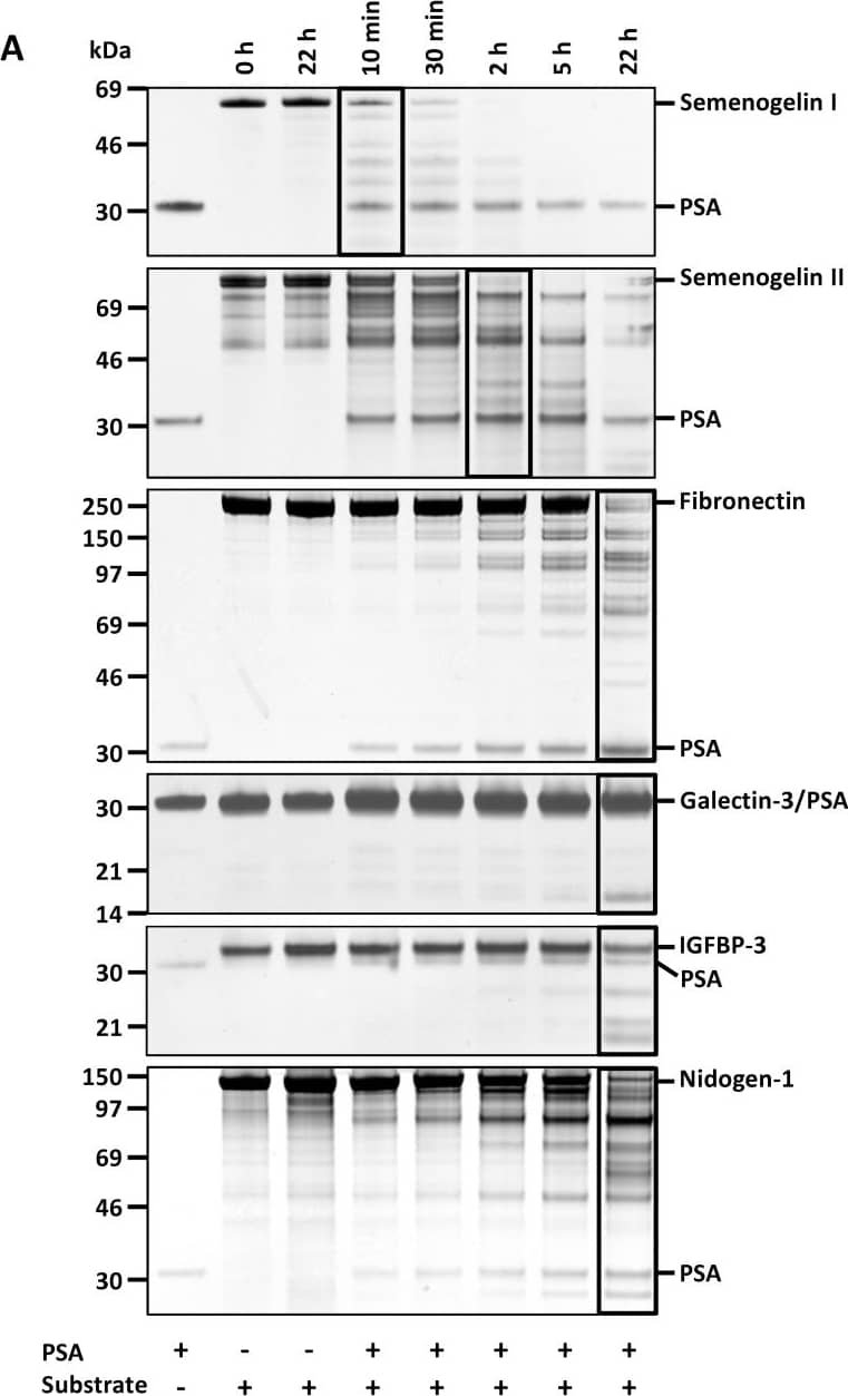
Detection of Human Nidogen-1/Entactin by Western Blot
Cleavage of protein substrates by PSA.(A) Characterization of the proteolytic activity of PSA (0.2 µM) during 22 h incubation towards different protein substrates (1 µM each, except 0.5 µM semenogelin I) by SDS-PAGE and silver staining. Approximate molecular weights of the proteins are: PSA (28 kDa), semenogelin I (50 kDa), semenogelin II (63 kDa), fibronectin (220 kDa), galectin-3 (26 kDa), IGFBP-3 (30 kDa) and nidogen-1 (130 kDa). The lanes in which ∼50% of the proteins were cleaved are bordered. (B) 1 µM MMP-3 (22 kDa), but not PSA, cleaved 1 µM plasminogen (88 kDa). Also 0.5 M fibronectin was incubated with 1 µM PSA as a control (SDS-PAGE with silver staining). Image collected and cropped by CiteAb from the following publication (https://dx.plos.org/10.1371/journal.pone.0107819), licensed under a CC-BY license. Not internally tested by R&D Systems.Applications for Human Nidogen-1/Entactin Antibody
Application
Recommended Usage
Immunohistochemistry
5-15 µg/mL
Sample: Immersion fixed paraffin-embedded sections of human heart
Sample: Immersion fixed paraffin-embedded sections of human heart
Western Blot
0.1 µg/mL
Sample: Recombinant Human Nidogen-1/Entactin (Catalog # 2570-ND)
Sample: Recombinant Human Nidogen-1/Entactin (Catalog # 2570-ND)
Reviewed Applications
Read 2 reviews rated 3.5 using AF2570 in the following applications:
Formulation, Preparation, and Storage
Purification
Antigen Affinity-purified
Reconstitution
Reconstitute at 0.2 mg/mL in sterile PBS. For liquid material, refer to CoA for concentration.
Formulation
Lyophilized from a 0.2 μm filtered solution in PBS with Trehalose. *Small pack size (SP) is supplied either lyophilized or as a 0.2 µm filtered solution in PBS.
Shipping
Lyophilized product is shipped at ambient temperature. Liquid small pack size (-SP) is shipped with polar packs. Upon receipt, store immediately at the temperature recommended below.
Stability & Storage
Use a manual defrost freezer and avoid repeated freeze-thaw cycles.
- 12 months from date of receipt, -20 to -70 °C as supplied.
- 1 month, 2 to 8 °C under sterile conditions after reconstitution.
- 6 months, -20 to -70 °C under sterile conditions after reconstitution.
Background: Nidogen-1/Entactin
Nidogen-1 (also entactin) is a 150 kDa, secreted, monomeric glycoprotein that serves as a major linking component of basement membranes (1-4). It is synthesized as a 1247 amino acid (aa) precursor with a 28 aa signal sequence and a 1219 aa mature protein. The molecule is modular in structure with five distinct regions. There are three globular domains (G1-3) separated by a mucin region and an extended rod-shaped segment (5-7). The N-terminal globular domain (G1) is 200 aa in length and seemingly unrelated to any known motif (8). The mucin region is nearly 160 aa in length and presumably O-glycosylated (2, 8). G2 and G3 are both approximately 300 aa in length. G2 is described as a Nidogen ( beta-barrel) domain, while C-terminal G3 assumes a beta-propeller configuration (1). The 250 aa rod-shaped segment has multiple EGF-like motifs and two thyroglobulin type 1 domains. Functionally, G1 is reported to bind type IV collagen (2, 7). The mucin region contains a short peptide that ligates alpha3 beta1 integrins (9, 10). G2 interacts with perlecan, and an RGD motif in the rod-shaped segment serves as a binding site for alphav beta3 integrins (9, 10). Finally, G3 is associated with laminin binding (2, 7). As a full-length molecule, the multiple extracellular matrix-binding sites of Nidogen-1 are well positioned to serve as anchor sites for basement membrane molecules. Nidogen-1 also undergoes proteolytic processing by at least two MMPs, MMP-7 and MMP-19 (10, 11). While this destroys the integrity of Nidogen-associated matrices, it also generates peptide fragments that are capable of inducing neutrophil chemotaxis and phagocytosis (10). Nidogen-2 is related to Nidogen-1 (≈ 50% aa identity) and shares many of the same adhesive properties as Nidogen-1 (12). Both bind perlecan plus collagens I and IV. Nidogen‑2, however, does not bind fibulin-1 or 2, and shows only modest interaction with laminin. Thus, although coexpressed, Nidogen-2 serves as only a partial substitute for Nidogen-1 (2, 12). Human Nidogen-1 shares 85% aa sequence identity with both mouse and rat Nidogen-1, and 88% aa sequence identity with canine Nidogen-1.
References
- Hohenester, E. and J. Engel (2002) Matrix Biol. 21:115.
- Miosge, N. et al. (2001) Histochem. J. 33:523.
- Charonis, A. et al. (2005) Curr. Med. Chem. 12:1495.
- Timpl, R. and J.C. Brown (1996) BioEssays 18:123.
- Nagayoshi, T. et al. (1989) DNA 8:581.
- Zimmerman, K. et al. (1995) Genomics 27:245.
- Fox, J.W. et al. (1991) EMBO J. 10:3137.
- Mayer, U. et al. (1995) Eur. J. Biochem. 227:681.
- Gresham, H.D. et al. (1996) J. Biol. Chem. 271:30587.
- Dong, L-J. et al. (1995) J. Biol. Chem. 270:15383.
- Titz, B. et al. (2004) Cell. Mol. Life Sci. 61:1826.
- Kohfeldt, K. et al. (1998) J. Mol. Biol. 282:99.
Alternate Names
Entactin-1, NID1, Nidogen1
Entrez Gene IDs
4811 (Human)
Gene Symbol
NID1
UniProt
Additional Nidogen-1/Entactin Products
Product Documents for Human Nidogen-1/Entactin Antibody
Product Specific Notices for Human Nidogen-1/Entactin Antibody
For research use only
Loading...
Loading...
Loading...
Loading...
Loading...