
Human MSP/MST1 Antibody
R&D Systems, part of Bio-Techne | Catalog # AF352

Scientific images may be resized, cropped, or adjusted for brightness or contrast for presentation purposes. These updates are limited to presentation and do not affect the underlying scientific interpretation of the data. Where available, additional source data may be provided upon request.
Conjugate
Catalog #
Key Product Details
Validated by
Biological Validation
Species Reactivity
Validated:
Human
Cited:
Human, Mouse
Applications
Validated:
Western Blot, Neutralization
Cited:
Immunohistochemistry-Paraffin, Western Blot, Neutralization, Flow Cytometry, Co-Immunoprecipitation
Label
Unconjugated
Antibody Source
Polyclonal Goat IgG
Product Specifications
Immunogen
Mouse myeloma cell line NS0-derived recombinant human MSP/MST1
Gln19-Gly711
Accession # AAA59872
Gln19-Gly711
Accession # AAA59872
Specificity
Detects human MSP/MST1 in direct ELISAs and Western blots. In direct ELISAs and Western blots, less than 5% cross‑reactivity with recombinant human HGF is observed.
Clonality
Polyclonal
Host
Goat
Isotype
IgG
Endotoxin Level
<0.10 EU per 1 μg of the antibody by the LAL method.
Scientific Data Images for Human MSP/MST1 Antibody
Chemotaxis Induced by MSP and Neutralization by Human MSP Antibody.
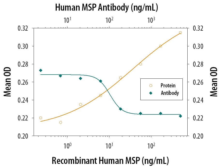
Recombinant Human MSP (Catalog # 352-MS) chemoattracts mouse peritoneal resident macrophages in a dose-dependent manner (orange line). The amount of cells that migrated through the filter was measured by LeukoStat™ staining (Fisher Scientific). Chemotaxis elicited by Recombinant Human MSP (50 ng/mL) is neutralized (green line) by increasing concentrations of Goat Anti-Human MSP Antigen Affinity-purified Polyclonal Antibody (Catalog # AF352). The ND50 is typically 0.05-0.2 µg/mL.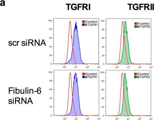
Detection of Mouse MSP/MST1 by Flow Cytometry
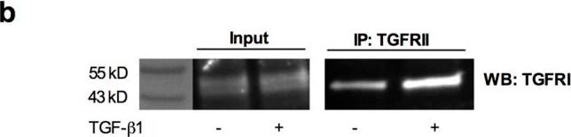
TGFRI and TGFRII association in fibulin-6 KD cells upon TGF-beta stimulus.scr siRNA or fibulin-6 siRNA transfected nCF, after TGF-beta stimulation are subjected to (a) FACS analysis. No difference in the amount of TGF betaRI and TGF betaRII on the surface of control and fibulin-6 KD cells was observed. (b) Immuno-precipitation of TGFRII from cell lysates of scr transfected and TGF-beta stimulated cells, shows more accumulation of TGFRI after western blotting compared to non TGF-beta treated nCF. (c) Immuno-precipitation of TGFRII followed by WB for TGFRI from cell lysates of scr transfected or fibulin-6 KD cells after TGF-beta stimulation display reduced association of receptors in fibulin-6 KD condition. The control input lanes shows no difference and IgG control is also clean (n = 4, p < 0.05). (d) Phosphorylation status of TGFRI was analyzed by western blot using phospho-TGFRI specific antibody. Densitometric analysis of western blot display decreased phosphorylation of TGFRI in fibulin-6 KD cells after TGF-beta stimulation compared to control cells (n = 5, p < 0.01, nonparametric Mann-Whitney U test). Image collected and cropped by CiteAb from the following open publication (https://pubmed.ncbi.nlm.nih.gov/28209981), licensed under a CC-BY license. Not internally tested by R&D Systems.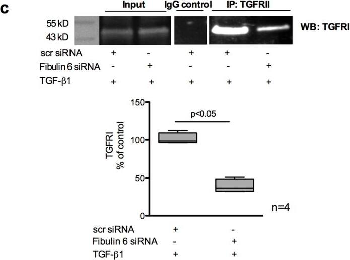
Detection of Mouse MSP/MST1 by Immunoprecipitation
TGFRI and TGFRII association in fibulin-6 KD cells upon TGF-beta stimulus.scr siRNA or fibulin-6 siRNA transfected nCF, after TGF-beta stimulation are subjected to (a) FACS analysis. No difference in the amount of TGF betaRI and TGF betaRII on the surface of control and fibulin-6 KD cells was observed. (b) Immuno-precipitation of TGFRII from cell lysates of scr transfected and TGF-beta stimulated cells, shows more accumulation of TGFRI after western blotting compared to non TGF-beta treated nCF. (c) Immuno-precipitation of TGFRII followed by WB for TGFRI from cell lysates of scr transfected or fibulin-6 KD cells after TGF-beta stimulation display reduced association of receptors in fibulin-6 KD condition. The control input lanes shows no difference and IgG control is also clean (n = 4, p < 0.05). (d) Phosphorylation status of TGFRI was analyzed by western blot using phospho-TGFRI specific antibody. Densitometric analysis of western blot display decreased phosphorylation of TGFRI in fibulin-6 KD cells after TGF-beta stimulation compared to control cells (n = 5, p < 0.01, nonparametric Mann-Whitney U test). Image collected and cropped by CiteAb from the following open publication (https://pubmed.ncbi.nlm.nih.gov/28209981), licensed under a CC-BY license. Not internally tested by R&D Systems.Applications for Human MSP/MST1 Antibody
Application
Recommended Usage
Western Blot
0.1 µg/mL
Sample: Recombinant Human MSP (Catalog # 352-MS)
Sample: Recombinant Human MSP (Catalog # 352-MS)
Neutralization
Measured by its ability to neutralize MSP-induced chemotaxis in mouse peritoneal resident macrophages. Ming-Hai, W. et al. (1994) J. Biol. Chem. 269:3436‑3440. The Neutralization Dose (ND50) is typically 0.05-0.2 µg/mL in the presence of 50 ng/mL Recombinant Human MSP.
Reviewed Applications
Read 1 review rated 4 using AF352 in the following applications:
Formulation, Preparation, and Storage
Purification
Antigen Affinity-purified
Reconstitution
Reconstitute at 0.2 mg/mL in sterile PBS. For liquid material, refer to CoA for concentration.
Formulation
Lyophilized from a 0.2 μm filtered solution in PBS with Trehalose. *Small pack size (SP) is supplied either lyophilized or as a 0.2 µm filtered solution in PBS.
Shipping
Lyophilized product is shipped at ambient temperature. Liquid small pack size (-SP) is shipped with polar packs. Upon receipt, store immediately at the temperature recommended below.
Stability & Storage
Use a manual defrost freezer and avoid repeated freeze-thaw cycles.
- 12 months from date of receipt, -20 to -70 °C as supplied.
- 1 month, 2 to 8 °C under sterile conditions after reconstitution.
- 6 months, -20 to -70 °C under sterile conditions after reconstitution.
Background: MSP/MST1
Macrophage stimulating protein (MSP), also known as HGF-like protein, and scatter factor-2, is a member of the HGF family of growth factors (1). MSP is secreted as an inactive single chain precursor (pro-MSP) that contains a PAN/APPLE-like domain, four kringle domains, and a peptidase S1 domain which lacks enzymatic activity (2). Human MSP shares 79% aa sequence identity with mouse MSP and 44% aa sequence identity with human HGF. Pro-MSP is secreted by hepatocytes under the positive and negative control of CBP in complex with either HNF-4 or RAR, respectively (3). Circulating pro-MSP is proteolytically cleaved in response to tissue injury to yield biologically active disulfide linked heterodimers consisting of a 45‑62 kDa alpha and a 25‑35 kDa beta chain (4, 5). Pro-MSP can be activated by MT-SP1, a transmembrane protease that is expressed on macrophages and is upregulated in many cancers (6). Heterodimeric MSP as well as the isolated beta chain bind to MSP R/Ron with high-affinity, although only heterodimeric MSP can induce receptor dimerization and signaling (7, 8). MSP induces macrophage and keratinocyte proliferation and osteoclast activation (9, 10). It also inhibits LPS- or IFN-induced iNOS and IL-12 expression by macrophages and prevents apoptosis of epithelial cells separated from the ECM (11, 12).
References
- Wang, M.-H. et al. (2002) Scand. J. Immunol. 56:545.
- Han, S. et al. (1991) Biochemistry 30:9768.
- Muraoka, R.S. et al. (1999) Endocrinology 140:187.
- Wang, M.-H. et al. (1996) J. Clin. Invest. 97:720.
- Nanney, L.B. et al. (1998) J. Invest. Dermatol. 111:573.
- Bhatt, A.S. et al. (2007) Proc. Natl. Acad. Sci. 104:5771.
- Wang, M.-H. et al. (1997) J. Biol. Chem. 272:16999.
- Danilkovitch, A. et al. (1999) J. Biol. Chem. 274:29937.
- Wang, M.-H. et al. (1996) Exp. Cell Res. 226:39.
- Kurihara, N. et al. (1998) Exp. Hematol. 26:1080.
- Morrison, A.C. et al. (2004) J. Immunol. 172:1825.
- Liu, Q.P. et al. (1999) J. Immunol. 163:6606.
Long Name
Macrophage Stimulating Protein/Macrophage Stimulating 1 [Hepatocyte Growth Factor-like]
Alternate Names
HGFL, MST1, SF2
Gene Symbol
MST1
UniProt
Additional MSP/MST1 Products
Product Documents for Human MSP/MST1 Antibody
Product Specific Notices for Human MSP/MST1 Antibody
For research use only
Loading...
Loading...
Loading...
Loading...
Loading...