
Human IL-33 Antibody
R&D Systems, part of Bio-Techne | Catalog # AF3625

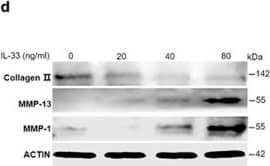

Scientific images may be resized, cropped, or adjusted for brightness or contrast for presentation purposes. These updates are limited to presentation and do not affect the underlying scientific interpretation of the data. Where available, additional source data may be provided upon request.
Conjugate
Catalog #
Key Product Details
Validated by
Orthogonal Validation
Species Reactivity
Validated:
Human
Cited:
Human, Mouse, Transgenic Mouse
Applications
Validated:
Immunohistochemistry, Western Blot, ELISA Capture (Matched Antibody Pair), Neutralization, Dual RNAscope ISH-IHC Compatible, Immunocytochemistry
Cited:
Immunohistochemistry, Immunohistochemistry-Paraffin, Immunohistochemistry-Frozen, Western Blot, Neutralization, Immunofluorescence, Immunocytochemistry, Cell Culture, ELISA Capture, ELISA Development
Label
Unconjugated
Antibody Source
Polyclonal Goat IgG
Product Specifications
Immunogen
E. coli-derived recombinant human IL-33
Ser112-Thr270
Accession # O95760
Ser112-Thr270
Accession # O95760
Specificity
Detects human IL-33 in ELISAs and Western blots. In sandwich ELISAs, less than 0.05% cross-reactivity with recombinant mouse (rm) IL‑33 is observed.
Clonality
Polyclonal
Host
Goat
Isotype
IgG
Endotoxin Level
<0.10 EU per 1 μg of the antibody by the LAL method.
Scientific Data Images for Human IL-33 Antibody
Cell Proliferation Induced by IL‑33 and Neutralization by Human IL‑33 Antibody.
Recombinant Human IL-33 (3625-IL) stimulates proliferation in the D10.G4.1 mouse helper T cell line in a dose-dependent manner (orange line). Under these conditions, proliferation elicited by Recombinant Human IL-33 (1 ng/mL) is neutralized (green line) by increasing concentrations of Goat Anti-Human IL-33 Antigen Affinity-purified Polyclonal Antibody (Catalog # AF3625). The ND50 is typically ≤ 3.0 µg/mL.
IL‑33 in Human Tonsil.
IL-33 was detected in immersion fixed paraffin-embedded sections of human tonsil using Goat Anti-Human IL-33 Antigen Affinity-purified Polyclonal Antibody (Catalog # AF3625) at 10 µg/mL overnight at 4 °C. Tissue was stained using the Anti-Goat HRP-DAB Cell & Tissue Staining Kit (brown; Catalog # CTS008) and counterstained with hematoxylin (blue). Specific staining was localized to nuclei. View our protocol for Chromogenic IHC Staining of Paraffin-embedded Tissue Sections.
IL-33 in Human Tonsil Using Dual RNAscope®ISH and IHC.
IL-33 mRNA was detected in formalin-fixed paraffin-embedded tissue sections of human tonsil probed with ACD RNAScope®Probe (Catalog # 400111) and stained using ACD RNAscope®2.5 HD Detection Reagents-Red (top image, Catalog # 32260). Adjacent tissue section was processed for immunohistochemistry using R&D Systems Goat Anti-Human IL-33 Antigen Affinity-purified Polyclonal Antibody (Catalog # AF3625) at 3 ug/mL with overnight incubation at 4 °C followed by incubation with the Anti-Goat IgG VisUCyte HRP Polymer Antibody (R&D Systems, Catalog # VC004) and DAB chromogen (lower image, yellow-brown). Tissues were counterstained with hematoxylin (blue).
Applications for Human IL-33 Antibody
Application
Recommended Usage
Dual RNAscope ISH-IHC Compatible
3-25 µg/mL
Sample: Immersion fixed paraffin-embedded sections of human tonsil
Sample: Immersion fixed paraffin-embedded sections of human tonsil
Immunocytochemistry
5-15 µg/mL
Sample: Immersion fixed human peripheral blood mononuclear cells treated with PMA and calcium ionomycin
Sample: Immersion fixed human peripheral blood mononuclear cells treated with PMA and calcium ionomycin
Immunohistochemistry
5-15 µg/mL
Sample: Immersion fixed paraffin-embedded sections of human tonsil
Sample: Immersion fixed paraffin-embedded sections of human tonsil
Western Blot
0.1 µg/mL
Sample: Recombinant Human IL-33 (Catalog # 3625-IL)
Sample: Recombinant Human IL-33 (Catalog # 3625-IL)
Neutralization
Measured by its ability to neutralize IL‑33-induced proliferation in the D10.G4.1 mouse helper T cell line. The Neutralization Dose (ND50) is ≤ 3.0 ug/mL in the presence of 1 ng/mL Recombinant Human IL-33 (Catalog # 3625-IL).
Human IL-33 Sandwich Immunoassay
Please Note: Optimal dilutions of this antibody should be experimentally determined.
Reviewed Applications
Read 8 reviews rated 4.6 using AF3625 in the following applications:
Formulation, Preparation, and Storage
Purification
Antigen Affinity-purified
Reconstitution
Reconstitute at 0.2 mg/mL in sterile PBS. For liquid material, refer to CoA for concentration.
Formulation
Lyophilized from a 0.2 μm filtered solution in PBS with Trehalose. See Certificate of Analysis for details.
*Small pack size (-SP) is supplied either lyophilized or as a 0.2 µm filtered solution in PBS.
*Small pack size (-SP) is supplied either lyophilized or as a 0.2 µm filtered solution in PBS.
Shipping
Lyophilized product is shipped at ambient temperature. Liquid small pack size (-SP) is shipped with polar packs. Upon receipt, store immediately at the temperature recommended below.
Stability & Storage
Use a manual defrost freezer and avoid repeated freeze-thaw cycles.
- 12 months from date of receipt, -20 to -70 °C as supplied.
- 1 month, 2 to 8 °C under sterile conditions after reconstitution.
- 6 months, -20 to -70 °C under sterile conditions after reconstitution.
Background: IL-33
IL-33, also known as NF-HEV and DVS 27, is a 30 kDa proinflammatory protein that may also regulate gene transcription (1‑3). DVS 27 was identifed as a gene that is up‑regulated in vasospastic cerebral arteries (1). NF-HEV was described as a nuclear factor that is preferentially expressed in the endothelial cells of high endothelial venules relative to endothelial cells from other tissues (2). IL-33 was identified based on sequence and structural homology with IL-1 family cytokines (3). DVS 27, NF-HEV, and IL-33 share 100% amino acid sequence identity. IL-33 is constitutively expressed in smooth muscle and airway epithelia. It is up‑regulated in arterial smooth muscle, dermal fibroblasts, and keratinocytes following IL-1 alpha or IL-1 beta stimulation (1, 3). Similar to IL-1, IL-33 can be cleaved in vitro by caspase-1, generating an N-terminal fragment that is slightly shorter than the C-terminal fragment (3, 4). The N-terminal portion of full length IL-33 contains a predicted bipartite nuclear localization sequence and a homeodomain-like helix-turn-helix DNA binding domain. By immunofluorescence, full length IL-33 localizes to the nucleus in HUVECs and transfectants (2). The C-terminal fragment, corresponding to mature IL-33, binds and triggers signaling through mast cell IL-1 R4/ST2L, a longtime orphan receptor involved in the augmentation of Th2 cell responses (3, 5-7). A ternary signaling complex is formed by the subsequent association of IL-33 and ST2L with IL-1R AcP (8). Stimulation of Th2 polarized lymphocytes with mature IL-33 in vitro induces IL-5 and IL-13 secretion (3). In vivo administration of mature IL-33 promotes increased production of IL-5, IL-13, IgE, and IgA, as well as splenomegaly and inflammatory infiltration of mucosal tissues (3). Full length and mature human IL-33 share 52‑58% aa sequence identity with mouse and rat IL-33. Human IL-33 shares less than 20% aa sequence identity with other IL-1 family proteins.
References
- Onda, H. et al. (1999) J. Cereb. Blood Flow Metab. 19:1279.
- Baekkevold, E.S. et al. (2003) Am. J. Pathol. 163:69.
- Schmitz, J. et al. (2005) Immunity 23:479.
- Black, R.A. et al. (1989) J. Biol. Chem. 264:5323.
- Xu, D. et al. (1998) J. Exp. Med. 187:787.
- Lohning, M. et al. (1998) Proc. Natl. Acad. Sci. USA 95:6930.
- Dinarello, C.A. (2005) Immunity 23:461.
- Chackerian, A.A. et al. (2007) J. Immunol. 179:2551.
Long Name
Interleukin 33
Alternate Names
C9orf26, DVS27, IL33, NF-HEV
Gene Symbol
IL33
UniProt
Additional IL-33 Products
Product Documents for Human IL-33 Antibody
Product Specific Notices for Human IL-33 Antibody
For research use only
Loading...
Loading...
Loading...
Loading...
Loading...
Loading...