
Human IL-1 RII APC-conjugated Antibody
R&D Systems, part of Bio-Techne | Catalog # FAB663A

Scientific images may be resized, cropped, or adjusted for brightness or contrast for presentation purposes. These updates are limited to presentation and do not affect the underlying scientific interpretation of the data. Where available, additional source data may be provided upon request.
Conjugate
Catalog #
Key Product Details
Validated by
Biological Validation
Species Reactivity
Validated:
Human
Cited:
Human
Applications
Validated:
Flow Cytometry
Cited:
Flow Cytometry
Label
Allophycocyanin (Excitation = 620-650 nm, Emission = 660-670 nm)
Antibody Source
Monoclonal Mouse IgG1 Clone # 34141
Product Specifications
Immunogen
S. frugiperda insect ovarian cell line Sf 21-derived recombinant human IL-1 RII
Phe14-Glu343 (Ser56Gly and Glu297Gly)
Accession # P27930
Phe14-Glu343 (Ser56Gly and Glu297Gly)
Accession # P27930
Specificity
Detects human IL-1 RII in direct ELSAs and Western blots. When used in combination with the biotinylated human IL-1 RII affinity purified polyclonal detection antibody (Catalog # BAF263) in sandwich ELISAs, no significant cross-reactivity or interference was observed with recombinant human (rh) IL-1ra, rhIL-1 RI, recombinant mouse IL-1ra, or recombinant rat IL-1ra.
Clonality
Monoclonal
Host
Mouse
Isotype
IgG1
Scientific Data Images for Human IL-1 RII APC-conjugated Antibody
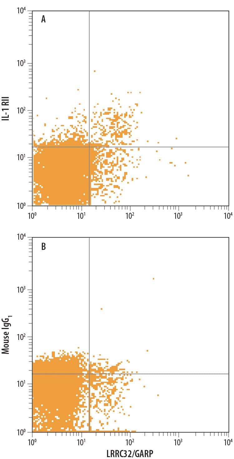
Detection of IL‑1 RII in Human PBMCs stimulated to induce Tregs by Flow Cytometry.
Human peripheral blood mononuclear cells (PBMCs), stimulated to induce Regulatory T Cells (Tregs) and gated on CD4+, were treated with 10 μg/mL Anti-CD3, 5 μg/mL Anti-CD28, 10 ng/mL Recombinant Human TGF-beta 1 (240-B), and 20 ng/mL Recombinant Human IL-2 (202-IL) for 48 hours and stained with Rat Anti-Human LRRC32/GARP PE-conjugated Monoclonal Antibody and either (A) Mouse Anti-Human IL-1 RII APC-conjugated Monoclonal Antibody (Catalog # FAB663A) or (B) Mouse IgG1Allophycocyanin Isotype Control (IC002A). View our protocol for Staining Membrane-associated Proteins.
Detection of IL-1 RII in HDLM-2 cells by Flow Cytometry
HDLM-2 cells were stained with Mouse Anti-Human IL-1 RII APC-conjugated Monoclonal Antibody (Catalog # FAB663A, filled histogram) or isotype control antibody (Catalog # IC002A, open histogram). View our protocol for Staining Membrane-associated Proteins.Applications for Human IL-1 RII APC-conjugated Antibody
Application
Recommended Usage
Flow Cytometry
10 µL/106 cells
Sample: Human peripheral blood mononuclear cells (PBMCs), stimulated to induce Regulatory T Cells (Tregs) and gated on CD4+, were treated with Anti-CD3, Anti-CD28, Recombinant Human TGF‑ beta1 (Catalog # 240-B), and Recombinant Human IL‑2 (Catalog # 202-IL)
Sample: Human peripheral blood mononuclear cells (PBMCs), stimulated to induce Regulatory T Cells (Tregs) and gated on CD4+, were treated with Anti-CD3, Anti-CD28, Recombinant Human TGF‑ beta1 (Catalog # 240-B), and Recombinant Human IL‑2 (Catalog # 202-IL)
Spectra Viewer
Plan Your Experiments
Use our spectra viewer to interactively plan your experiments, assessing multiplexing options. View the excitation and emission spectra for our fluorescent dye range and other commonly used dyes.
Spectra Viewer
Formulation, Preparation, and Storage
Purification
Protein A or G purified from ascites
Formulation
Supplied in a saline solution containing BSA and Sodium Azide.
Shipping
The product is shipped with polar packs. Upon receipt, store it immediately at the temperature recommended below.
Stability & Storage
Protect from light. Do not freeze.
- 12 months from date of receipt, 2 to 8 °C as supplied.
Background: IL-1 RII
Two distinct types of receptors that bind the pleiotropic cytokines IL-1 alpha and IL-1 beta have been described. The IL-1 receptor type I is an 80 kDa transmembrane protein that is expressed predominantly by T cells, fibroblasts, and endothelial cells. IL-1 receptor type II is a 68 kDa transmembrane protein found on B lymphocytes, neutrophils, monocytes, large granular leukocytes, and endothelial cells. Both receptors are members of the immunoglobulin superfamily and show approximately 28% sequence similarity in their extracellular domains. The two receptor types do not heterodimerize in a receptor complex. An IL-1 receptor accessory protein that can heterodimerize with the type I receptor in the presence of IL-1 alpha or IL-1 beta, but not IL-1ra, was identified (1). This type I receptor complex appears to mediate all the known IL-1 biological responses. The receptor type II has a short cytoplasmic domain and does not transduce IL-1 signals. In addition to the membrane-bound form of IL-1 RII, a naturally-occurring soluble form of IL-1 RII has been described. It has been suggested that the type II receptor, either as the membrane-bound or as the soluble form, serves as a decoy for IL-1 and inhibits IL-1 action by blocking the binding of IL-1 to the signaling type I receptor complex. Recombinant IL-1 soluble receptor type II is a potent antagonist of IL-1 action.
References
- Greenfeder, S. et al. (1995) J. Biol. Chem. 270:13757.
Long Name
Interleukin 1 Receptor II
Alternate Names
CD121b, IL-1 R beta, IL-1RII, IL1R2, IL1RII
Gene Symbol
IL1R2
UniProt
Additional IL-1 RII Products
Product Documents for Human IL-1 RII APC-conjugated Antibody
Product Specific Notices for Human IL-1 RII APC-conjugated Antibody
For research use only
Loading...
Loading...
Loading...
Loading...
Loading...