
Glut1 Antibody (MSVA-401R)
Novus Biologicals, part of Bio-Techne | Catalog # NBP3-57191
Recombinant Monoclonal Antibody

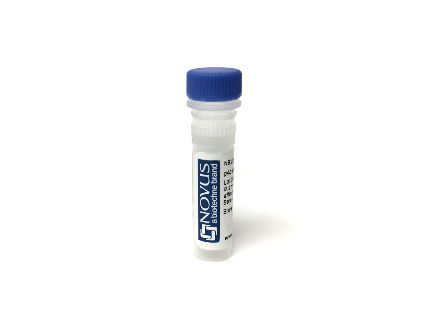
Key Product Details
Species Reactivity
Human
Applications
Immunohistochemistry, Immunohistochemistry-Paraffin
Label
Unconjugated
Antibody Source
Monoclonal Rabbit IgG Clone # MSVA-401R
Concentration
Please see the vial label for concentration. If unlisted please contact technical services.
Product Specifications
Immunogen
Recombinant fragment of human GLUT1 protein (around aa 203-305) (exact sequence is proprietary) Swissprot P11166
Localization
Cell Surface
Clonality
Monoclonal
Host
Rabbit
Isotype
IgG
Description
This antibody is highly specific and optimized for use in Immunohistochemistry.
It has been validated on TMAs covering 76 normal tissues and numerous cancer tissues.
The antibody concentration is optimized for dilution within dilution range using commercially available antibody diluent for immunohistochemistry.
Positive Control
Postive control:Brain: A strong GLUT1 staining of endothelial cells should be seen..
Negative Control
Negitve control:Brain: GLUT1 staining should be absent in all cells/structures except blood vessels.
Storage
Antibody can be stored at 4degC. For extended storage, aliquot and store at -20degC. Avoid repeated freeze-thaw cycles.
Applications for Glut1 Antibody (MSVA-401R)
Application
Recommended Usage
Immunohistochemistry-Paraffin
1:100 1:200
Application Notes
Manual Protocol: Freshly cut sections should be used (less than 10 days between cutting and staining). Heat-induced antigen retrieval for 5 minutes in an autoclave at 121C in pH 7,8 Target Retrieval Solution buffer. Apply the antibody at a dilution of 1:150 at 37C for 60 minutes. Visualization of bound antibody by the EnVision Kit (Dako, Agilent) according to the manufacturer's directions.
Formulation, Preparation, and Storage
Purification
Protein A or G purified
Formulation
10mM PBS
Preservative
0.1% Sodium Azide
Concentration
Please see the vial label for concentration. If unlisted please contact technical services.
Shipping
The product is shipped with polar packs. Upon receipt, store it immediately at the temperature recommended below.
Stability & Storage
Store at 4C.
Background: Glut1
GLUT1 (Human glycosylated form theoretical molecular weight 55kDa) functions primarily as a glucose transporter but can transport other substrates including mannose, galactose and glucosamine across the membrane (3). Like other GLUT family members, GLUT1 is broadly expressed, nevertheless it is the predominant glucose transporter expressed in red blood cells and brain endothelial cells (1). SLC2A1 mutations underscore the autosomal dominant disorder GLUT1 deficiency syndrome (GLUTI-DS) which is characterized by low glucose levels in the brain or hypoglycorrhachia due to insufficient glucose transport across the blood brain barrier (2, 4, 5). Phenotypically, GLUT1-DS is characterized by early onset seizures, neurologic developmental delay, microcephaly, and ataxia (4). GLUT1 is highly expressed in the endothelium of cutaneous vascular lesions and serves as a marker for the diagnosis of juvenile or infantile hemangiomas (6).
References
1. Augustin, R. (2010). The protein family of glucose transport facilitators: It's not only about glucose after all. IUBMB Life. https://doi.org/10.1002/iub.315
2. Mueckler, M., & Thorens, B. (2013). The SLC2 (GLUT) family of membrane transporters. Molecular Aspects of Medicine. https://doi.org/10.1016/j.mam.2012.07.001
3. Stein, W. D., & Litman, T. (2015). Carrier-Mediated Transport. In Channels, Carriers, and Pumps. https://doi.org/10.1016/b978-0-12-416579-3.00004-6
4. Pearson, T. S., Akman, C., Hinton, V. J., Engelstad, K., & De Vivo, D. C. (2013). Phenotypic spectrum of glucose transporter type 1 deficiency syndrome (Glut1 DS). Current Neurology and Neuroscience Reports. https://doi.org/10.1007/s11910-013-0342-7
5. Messana, T., Russo, A., Vergaro, R., Boni, A., Santucci, M., & Pini, A. (2018). Glucose transporter type 1 deficiency syndrome: Developmental delay and early-onset ataxia in a novel mutation of the SLC2A1 gene. Journal of Pediatric Neurosciences. https://doi.org/10.4103/JPN.JPN_169_17
6. van Vugt, L. J., van der Vleuten, C. J. M., Flucke, U., & Blokx, W. A. M. (2017). The utility of GLUT1 as a diagnostic marker in cutaneous vascular anomalies: A review of literature and recommendations for daily practice. Pathology Research and Practice. https://doi.org/10.1016/j.prp.2017.04.023
Long Name
Glucose Transporter Type 1
Alternate Names
DYT17, DYT18, DYT9, EIG12, GLUT1DS, SLC2A1
Gene Symbol
SLC2A1
Additional Glut1 Products
Product Specific Notices for Glut1 Antibody (MSVA-401R)
This product is for research use only and is not approved for use in humans or in clinical diagnosis. Primary Antibodies are guaranteed for 1 year from date of receipt.
Loading...
Loading...
Loading...
Loading...