
53BP1 Antibody [DyLight 550]
Novus Biologicals, part of Bio-Techne | Catalog # NB100-304R

![53BP1 Antibody [DyLight 550]](https://resources.bio-techne.com/images/products/nb100-304r_rabbit-polyclonal-53bp1-antibody-dylight-550-immunocytochemistry-immunofluorescence-266202582231..jpg "53BP1 in SJCRH30 Human Cell Line.")
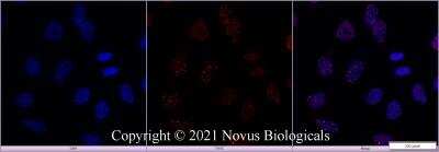
Scientific images may be resized, cropped, or adjusted for brightness or contrast for presentation purposes. These updates are limited to presentation and do not affect the underlying scientific interpretation of the data. Where available, additional source data may be provided upon request.
Conjugate
Catalog #
Forumulation
Catalog #
Key Product Details
Species Reactivity
Validated:
Human, Mouse, Rat
Cited:
Mouse
Predicted:
Chimpanzee (96%), Feline (90%), Orangutan (96%), Porcine (90%), Rabbit (90%), Sheep (90%). Backed by our 100% Guarantee.
Applications
Validated:
Knockout Validated, Immunohistochemistry, Immunohistochemistry-Paraffin, Immunohistochemistry-Frozen, Western Blot, Immunoblotting, Flow Cytometry, Flow (Intracellular), Immunocytochemistry/ Immunofluorescence, Immunoprecipitation, Chromatin Immunoprecipitation, Chromatin Immunoprecipitation (ChIP), In-situ Hybridization, Knockdown Validated
Cited:
Immunohistochemistry-Paraffin
Label
DyLight 550 (Excitation = 562 nm, Emission = 576 nm)
Antibody Source
Polyclonal Rabbit IgG
Concentration
Please see the vial label for concentration. If unlisted please contact technical services.
Product Specifications
Immunogen
Partial synthetic peptide made to an internal portion of human 53BP1 (between amino acids 350-400) [NP_005648.1].
Reactivity Notes
Predicted cross-reactivity based on sequence identity: Gorilla (96%), Gibbon (94%), Marmoset (92%).
Marker
DNA Double Strand Break Marker
Clonality
Polyclonal
Host
Rabbit
Isotype
IgG
Description
This conjugate is made on demand and is not held in inventory. Each order represents a unique lot. All conjugates are generated using validated conjugation protocols and meet strict degree of labeling (F/P) specifications; additional information is available upon request. The antibody concentration is provided on the individual product label.
For specialized conjugation needs, like large quantities, specific concentrations, and defined F/P ratios, bulk and custom antibody conjugation services are available.
For specialized conjugation needs, like large quantities, specific concentrations, and defined F/P ratios, bulk and custom antibody conjugation services are available.
Scientific Data Images for 53BP1 Antibody [DyLight 550]
Staining of 53BP1 in HeLa Cells Using Conjugated 53BP1 Antibody
HeLa cells were fixed in 4% paraformaldehyde for 10 minutes and permeabilized in 0.5% Triton X-100 in PBS for 5 minutes. The cells were incubated with anti-53BP1 Antibody conjugated to DyLight 550 (NB100-304R) at 5 ug/ml for 1 hour at room temperature. Nuclei were counterstained with DAPI (Blue). Cells were imaged using a 100X objective and digitally deconvolved.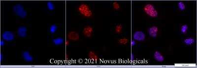
Imaging of HeLa Cells Stained for 53BP1 Using Conjugated 53BP1 Antibody
HeLa cells were fixed in 4% paraformaldehyde for 10 minutes and permeabilized in 0.5% Triton X-100 in PBS for 5 minutes. The cells were incubated with 53BP1 Antibody conjugated to DyLight 550 (NB100-304R) at 5 ug/ml for 1 hour at room temperature. Nuclei were counterstained with DAPI (Blue). Cells were imaged using a 40X objective.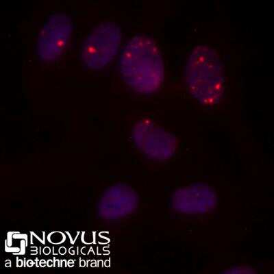
Immunocytochemistry/Immunofluorescence Staining of 53BP1 in HeLa Cells Using Conjugated 53BP1 Antibody
HeLa cells were fixed for 10 minutes using 10% formalin and then permeabilized for 5 minutes using 1X PBS + 0.5% Triton-X100. The cells were incubated with 53BP1 Antibody conjugated to DyLight 550 (Catalog #NB100-304R) at 10ug/ml for 1 hour at room temperature. Nuclei were counterstained with DAPI (Blue). Cells were imaged using a 40X objective.Applications for 53BP1 Antibody [DyLight 550]
Application
Recommended Usage
Chromatin Immunoprecipitation
Optimal dilutions of this antibody should be experimentally determined.
Chromatin Immunoprecipitation (ChIP)
Optimal dilutions of this antibody should be experimentally determined.
Flow (Intracellular)
Optimal dilutions of this antibody should be experimentally determined.
Flow Cytometry
Optimal dilutions of this antibody should be experimentally determined.
Immunoblotting
Optimal dilutions of this antibody should be experimentally determined.
Immunocytochemistry/ Immunofluorescence
Optimal dilutions of this antibody should be experimentally determined.
Immunohistochemistry
Optimal dilutions of this antibody should be experimentally determined.
Immunohistochemistry-Frozen
Optimal dilutions of this antibody should be experimentally determined.
Immunohistochemistry-Paraffin
Optimal dilutions of this antibody should be experimentally determined.
Immunoprecipitation
Optimal dilutions of this antibody should be experimentally determined.
In-situ Hybridization
Optimal dilutions of this antibody should be experimentally determined.
Knockdown Validated
Optimal dilutions of this antibody should be experimentally determined.
Knockout Validated
Optimal dilutions of this antibody should be experimentally determined.
Western Blot
Optimal dilutions of this antibody should be experimentally determined.
Application Notes
Recommended applications are based on validated applications from the unconjugated base product NB100-304. This conjugated antibody is not kept in inventory and is made to order using validated conjugation protocols.
Spectra Viewer
Plan Your Experiments
Use our spectra viewer to interactively plan your experiments, assessing multiplexing options. View the excitation and emission spectra for our fluorescent dye range and other commonly used dyes.
Spectra Viewer
Formulation, Preparation, and Storage
Purification
Immunogen affinity purified
Formulation
50mM Sodium Borate
Preservative
0.05% Sodium Azide
Concentration
Please see the vial label for concentration. If unlisted please contact technical services.
Shipping
The product is shipped with polar packs. Upon receipt, store it immediately at the temperature recommended below.
Stability & Storage
Store at 4C in the dark.
Background: 53BP1
References
1.Henry, E., Souissi-Sahraoui, I., Deynoux, M., Lefevre, A., Barroca, V., Campalans, A., . . . Arcangeli, M. L. (2019). Human hematopoietic stem/progenitor cells display ROS-dependent long-term hematopoietic defects after exposure to low dose of ionizing radiations. Haematologica. doi:10.3324/haematol.2019.226936
2.Janoshazi, A. K., Horton, J. K., Zhao, M. L., Prasad, R., Scappini, E. L., Tucker, C. J., & Wilson, S. H. (2020). Shining light on the response to repair intermediates in DNA of living cells. DNA Repair (Amst), 85, 102749. doi:10.1016/j.dnarep.2019.102749
Long Name
p53 Binding Protein 1
Alternate Names
p202, TP53BP1
Gene Symbol
TP53BP1
Additional 53BP1 Products
Product Documents for 53BP1 Antibody [DyLight 550]
Product Specific Notices for 53BP1 Antibody [DyLight 550]
DyLight (R) is a trademark of Thermo Fisher Scientific Inc. and its subsidiaries.
This product is for research use only and is not approved for use in humans or in clinical diagnosis. Primary Antibodies are guaranteed for 1 year from date of receipt.
Loading...
Loading...
Loading...
Loading...
Loading...
Loading...