Why Visualize RNA and Protein Together?
Understanding the spatial relationship between RNA and protein is crucial for gaining a holistic view of cellular processes. However, analyzing these molecules separately, as has been done traditionally, misses the intricate interplay that defines tissue function and disease progression. Spatial Multiomics examines RNA and proteins in relation to tissue morphology enabling a complete understanding of gene expression regulation and protein function within specific cellular contexts.
Discover Spatial Multiomic Insights
Secreted Protein and its Origin
Secreted Protein and its Origin
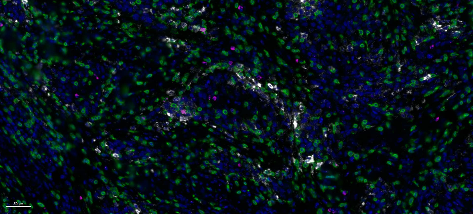
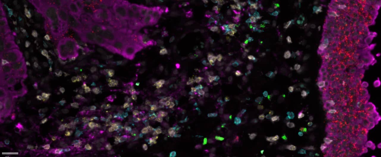
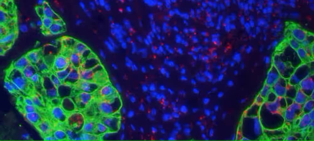
RNAscope Multiplex V2 assay for studying Cytotoxic T lymphocytes in cervical cancer tissue. T cells were identified using protein marker CD3 while cytokine markers IFNG and CXCL9 were detected using RNA.
Characterize Immune Cells in the Tumor Microenvironment
Characterize Immune Cells in the Tumor Microenvironment
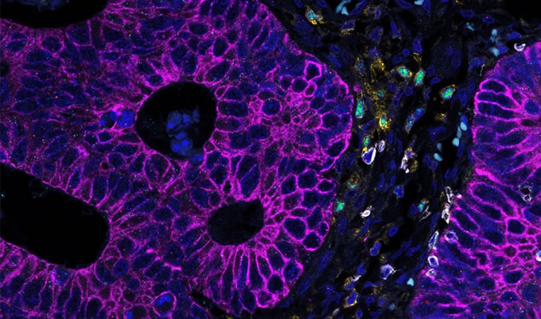
Visualizing TIL activation in Colon cancer. Using codetection to identify tumor infiltrating regulatory T cells marked by CD4+/FOXP3+ , Cytotoxic T lymphocytes seen as CD8+/ IFNG+.
Visualize Changes in Cell State
Visualize Changes in Cell State
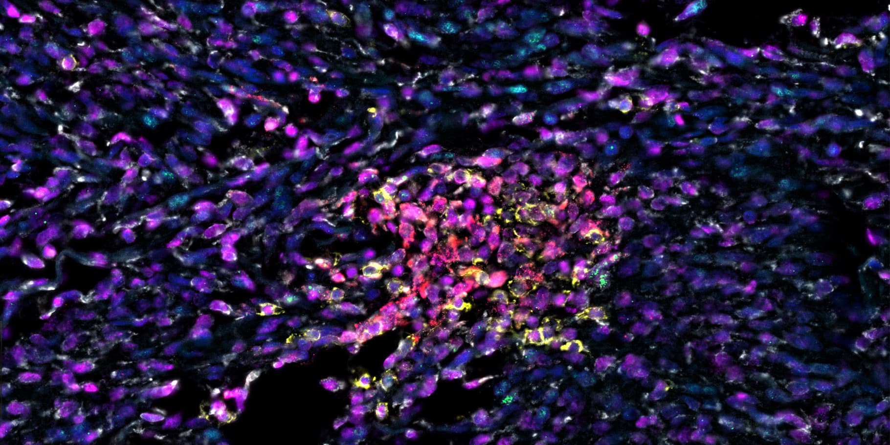
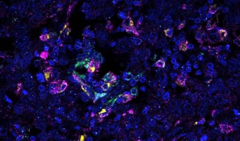
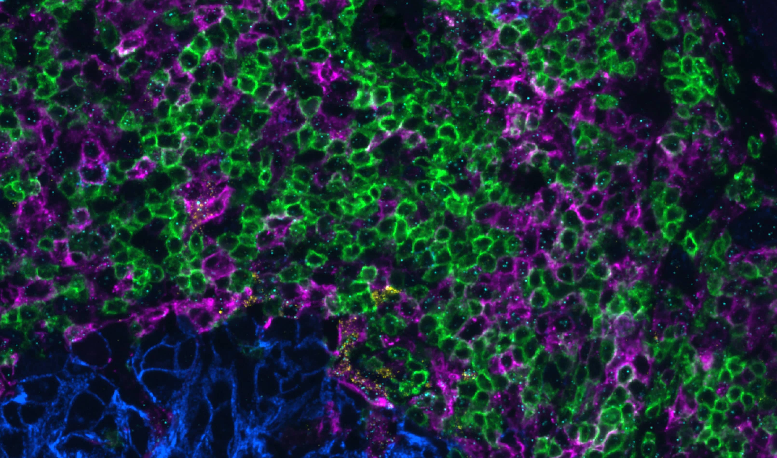
Mapping tumor associated M1/M2 macrophages marked by CD163+ / CD68+ , IL1B+ / IL10+ in cervical cancer tissue
Reveal Functional Protein-Protein Interactions
Reveal Functional Protein-Protein Interactions
ProximityScope assay reveals heterodimerization of receptors on tumor cells in head and neck cancer by visualizing EGFR-HER2 proximity.
Decode Neurodegeneration
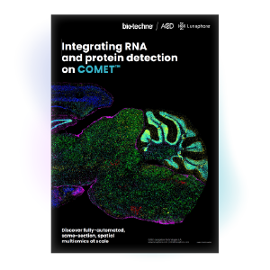
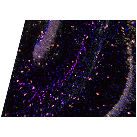
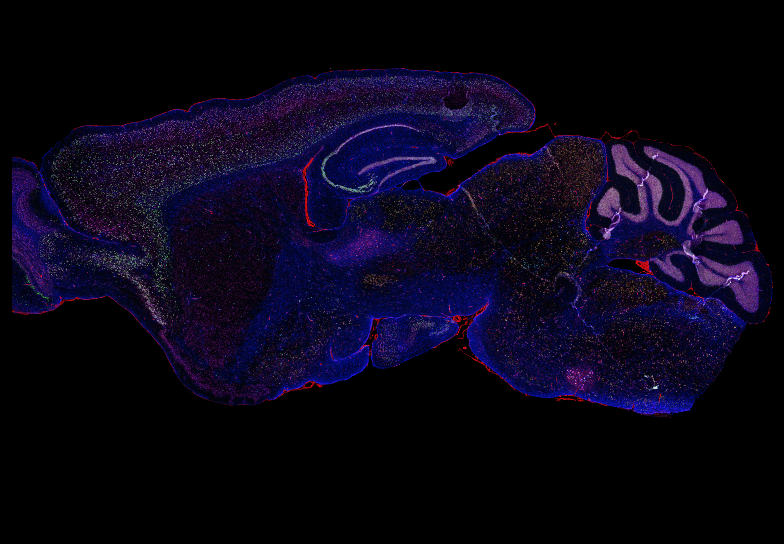
Decode Neurodegeneration
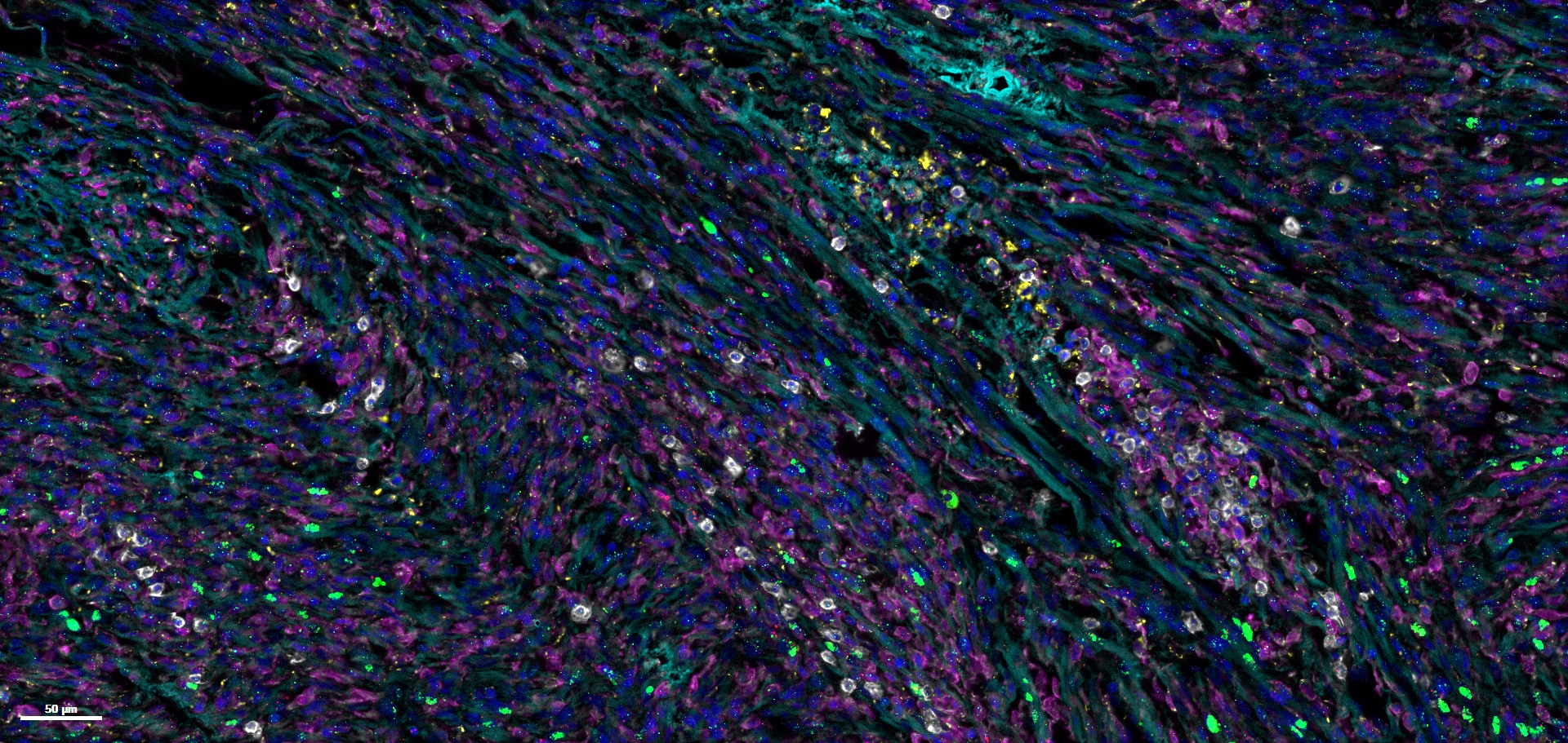
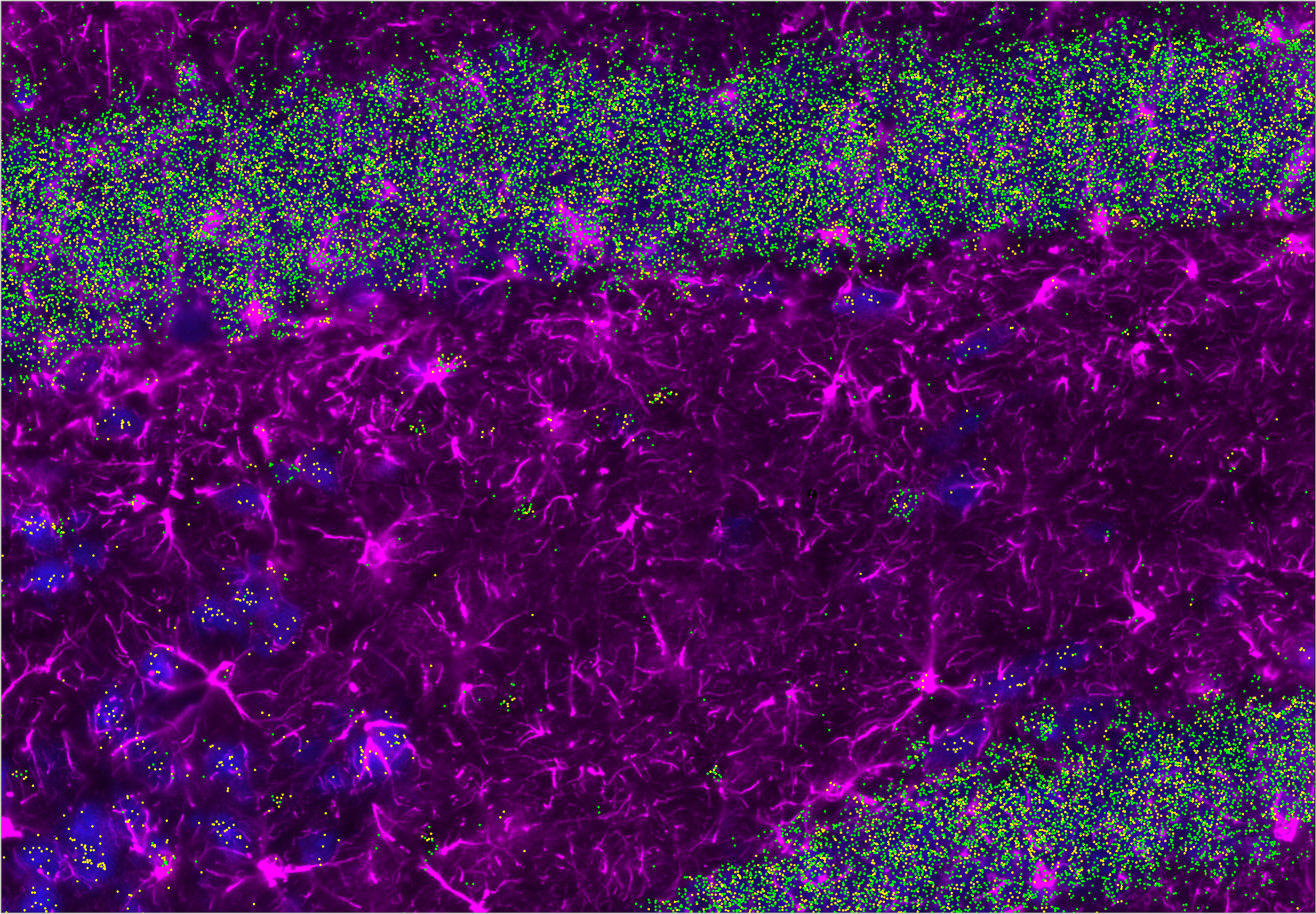
Identify neuronal and glial activation states through same-section spatial multiomics. RNA–protein co-detection on COMET, combined with HORIZON analysis, delivers confident, quantitative insight from a single tissue section.
Uncover Functional Immune States in Cancer
Uncover Functional Immune States in Cancer
Link immune phenotypes to cytokine expression using same-section spatial multiomics. Use COMET to enable precise mapping of immune activation and suppression within tumors.
- Comprehensive Insights: Capture the full picture of cellular activities
- Enhanced Data Quality: Achieve high-resolution, colocalized detection
- Improved Research Outcomes: Uncover new therapeutic targets and disease outcomes
RNAscope Multiomic LS Assay
RNAscope Multiomic LS Assay
Proven class-leading single-molecule RNAscope technology is leveraged to enable simultaneous detection of up to 6 total proteins and RNA targets with single cell resolution automated on the automated assay for the BOND RX™ by Leica Biosystems.
RNAscope HiPlex Pro for COMET
RNAscope HiPlex Pro for COMET
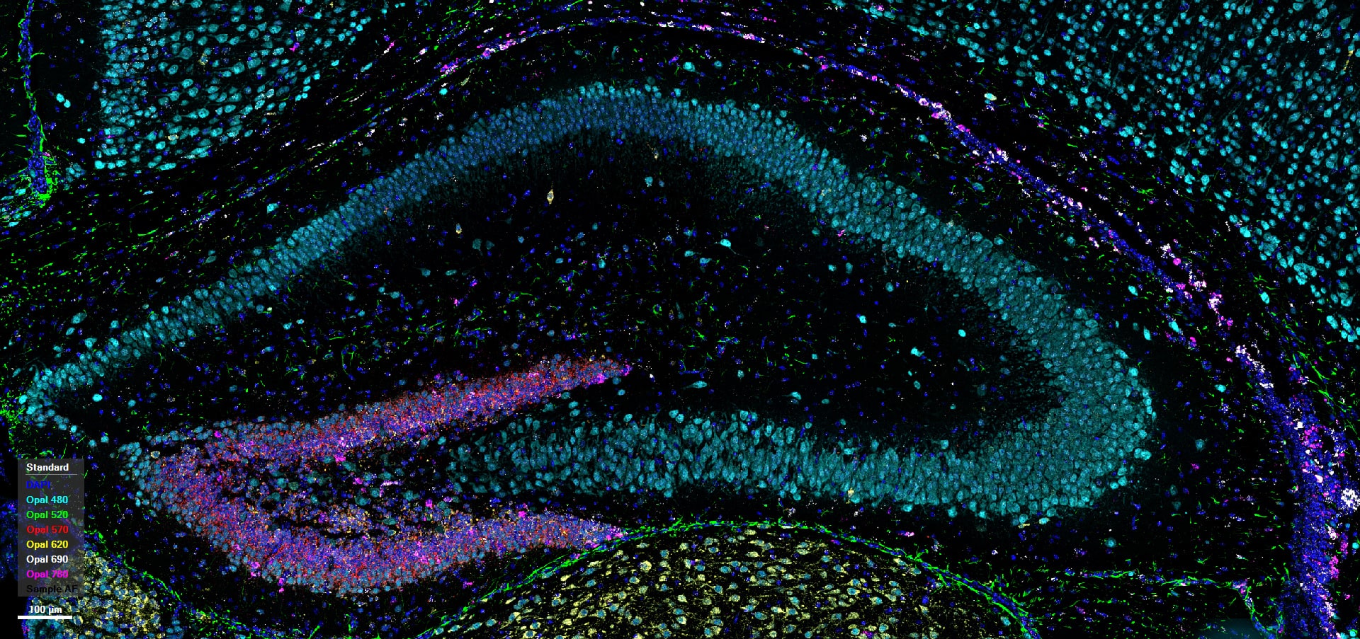
Combining RNAscope HiPlex Pro with seqIF (sequential immunofluorescence) on COMET platform with a protease-free, fully automated workflow, same-section RNA and multiplex protein detection with the ability to select any 12 RNAscope targets, and up to 24 IF targets using off-the-shelf, non-conjugated primary antibodies.
Protease-free Sequential RNA-Protein Detection
Protease-free Sequential RNA-Protein Detection
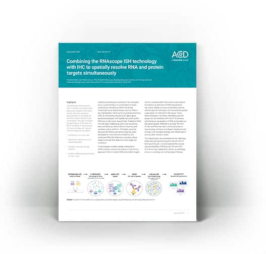
Leverage the robustness of RNAscope technology by combining in situ hybridization and IHC protocol in the same sample slide leveraging the sequential workflow for both manual and automated platforms from both Leica Biosystems and Roche Tissue Diagnostics.
Customer Success Stories
UCSF School of Medicine

Heidelberg University Hospital
Webinar: Expand your Multiomic Capabilities with RNAscope
In this webinar we provide an overview of RNAscope ISH technology, and the new RNAscope Multiomic LS assay. Learn about the extensive menu of RNAscope assays available that will enable you to detect a wide range of different RNA biomarkers in any species or tissue. Perform true same-slide multiomics using our new 6-plex capable RNAscope Multiomic kit leveraging a protease-free workflow that delivers staining performance, preserved tissue morphology, and simplified assay design.