
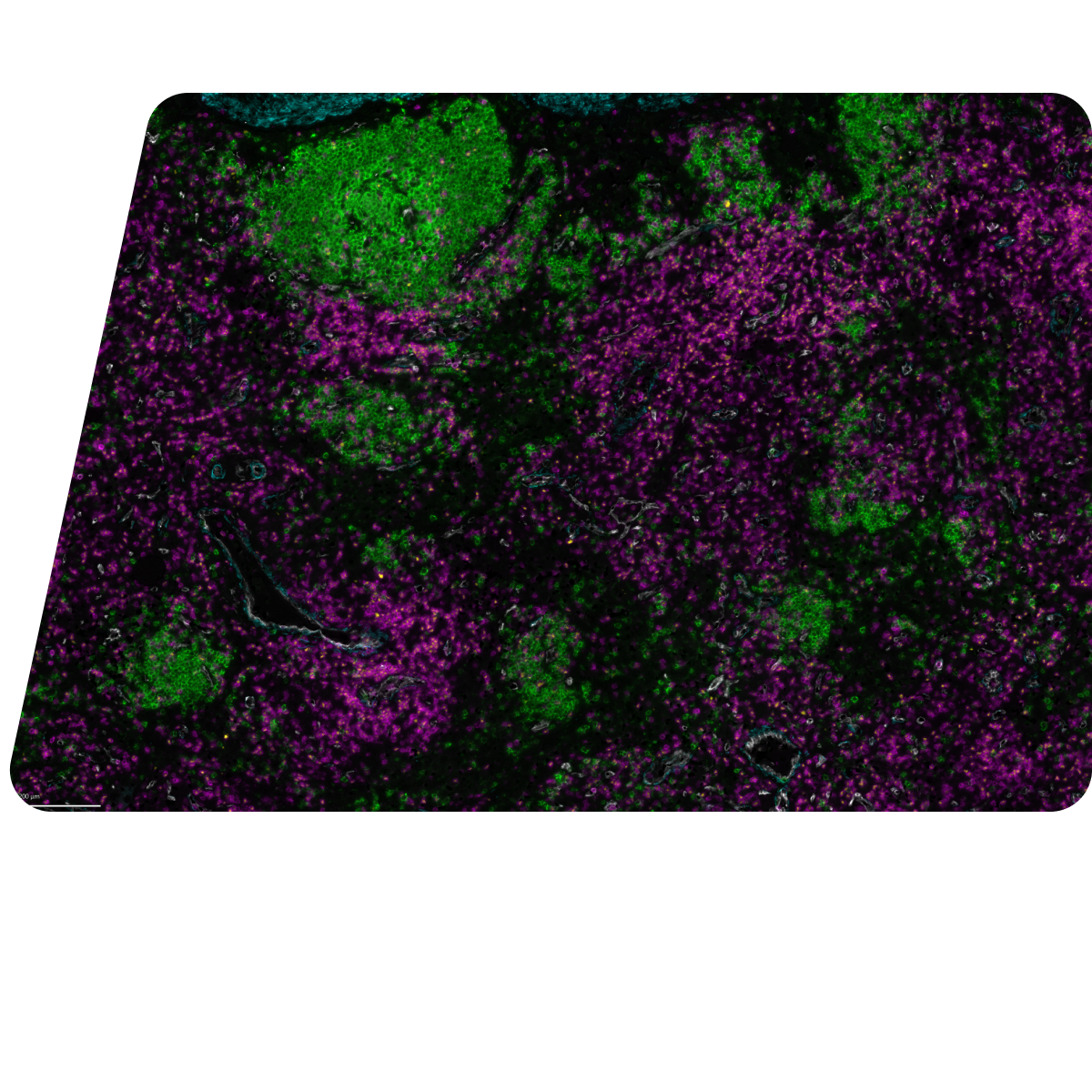
Beyond Tissue Morphology: Spatial Phenotyping in Hematological Malignancies
Resolve the cellular architecture of hematological malignancies with end-to-end spatial proteomics and multiomics on COMET™.
Characterise immune organisation, tumour niches and bone marrow architecture to uncover spatially informed biomarkers. COMET™ maximises the insight generated from limited tissue while preserving its morphological context.
The ASH Annual Meeting brings together the latest scientific and clinical advances across classical and malignant hematology, with a strong focus on translating research into better patient care.
Find us at booth #700 and bring us your most challenging translational question.
Discover COMET™ at ASH 2026
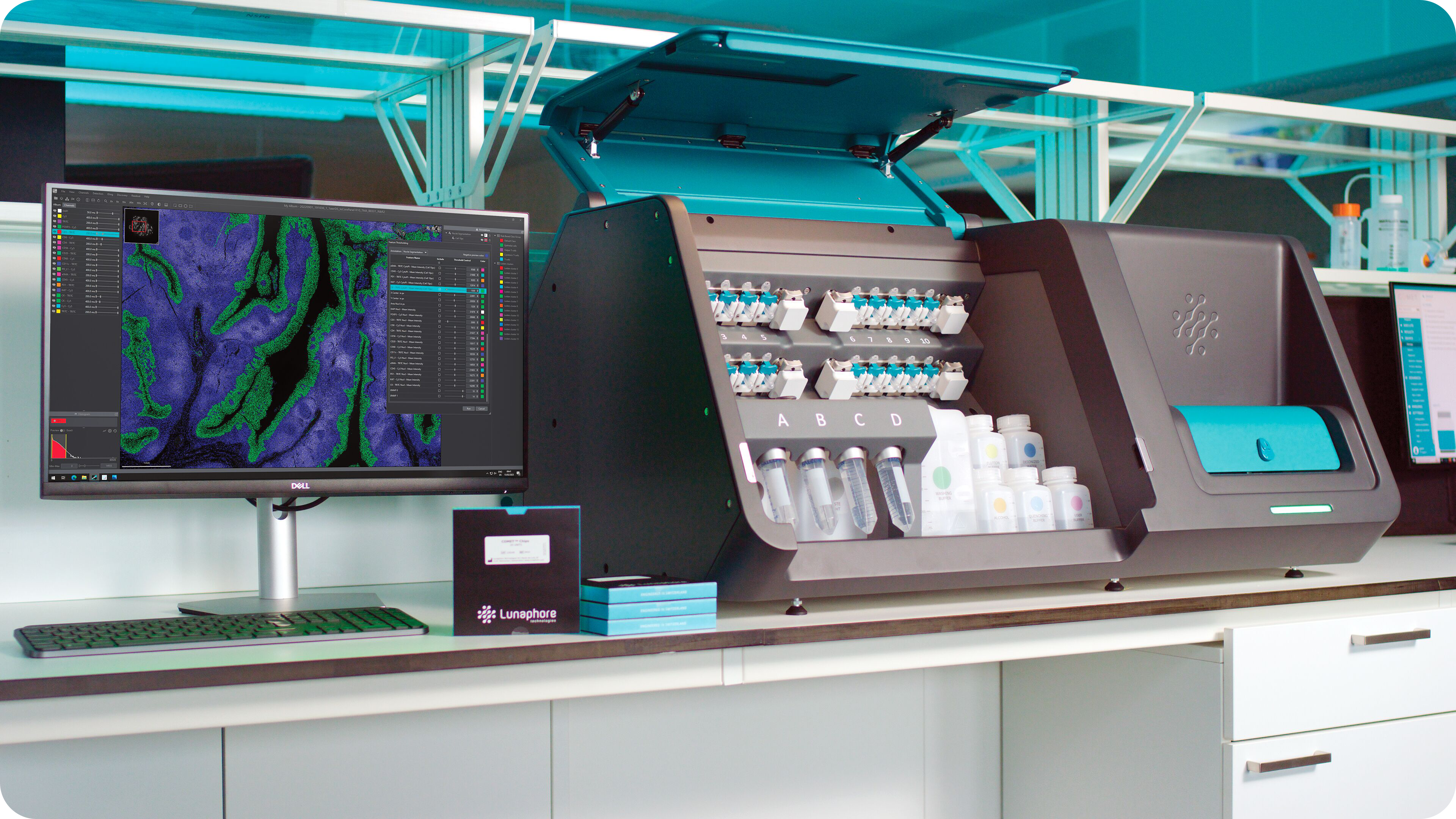
COMET™ is a fully-automated spatial biology platform designed to support multiplex protein analysis and spatial multiomics research across entire tissue sections.
The platform integrates staining, image acquisition and image preprocessing, helping researchers move from sample to spatially resolved data through a consistent and robust workflow. COMET™ supports off-the-shelf, label-free primary antibodies, allowing researchers to apply existing IHC-validated and clinically approved antibody clones without the need of upstream antibody conjugation or barcoding.
Product highlights
- Automated staining, imaging and image preprocessing
- Precision microfluidic technology
- Off-the-shelf, label-free primary antibodies
- Flexible and adaptable panel development
- Preserved tissue morphology and epitope stability
- High-plex same-section RNA and protein analysis
- Research applications across discovery, translational research and biomarker development
Spatial Evidence for The Translational Pathway
Make every sample count
Limited biopsy material requires technologies that generate high-content information without compromising tissue integrity.
COMET™ enables automated, highly multiplexed spatial protein analysis from a single tissue section. Precision microfluidics, integrated staining and imaging, and the use of off-the-shelf primary antibodies make it possible to study complex cell phenotypes and tissue structures while preserving morphology and epitopes for downstream applications.

Preserve limited samples
Generate high-plex spatial proteomics and multiomics data from a single tissue section, while maintaining tissue morphology.

Standardize complex workflows
Automate staining, image acquisition and image preprocessing to reduce manual variability and hands-on time.

Connect biology to treatment response
Resolve cell phenotypes, cellular neighbourhoods and tissue structures associated with disease progression and therapeutic response.

Design flexible panels
Use standard, label-free primary antibodies without requiring antibody conjugation or barcoding.
Bring Your Most Challenging Hematology Samples to ASH
Tell us about your tissue, markers and translational objective. Our spatial biology team can help you explore an appropriate COMET™ workflow and identify the most valuable next step for your research.
Meet with the Bio-Techne Spatial team to:
- Discuss a translational study design or biomarker hypothesis.
- Explore a protein or multiomics panel design strategy.
- Review the suitability of your tissue type and cohort.
- Request a tailored COMET™ demonstration.