
Mouse IgG2A Antibody
R&D Systems, part of Bio-Techne | Catalog # MAB0033
Recombinant Monoclonal Antibody.

Key Product Details
Species Reactivity
Mouse
Applications
Flow Cytometry, Western Blot
Label
Unconjugated
Antibody Source
Monoclonal Rabbit IgG Clone # 2214A
Product Specifications
Immunogen
Mouse IgG
Specificity
Detects mouse IgG
Clonality
Monoclonal
Host
Rabbit
Isotype
IgG
Scientific Data Images for Mouse IgG2A Antibody
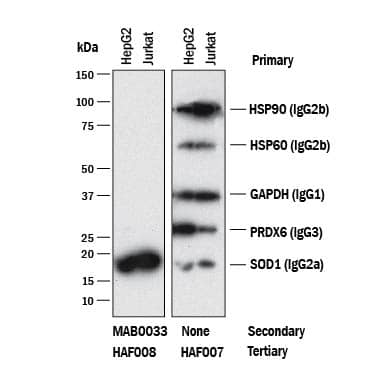
Detection of Mouse IgG2APrimary Antibodies by Western Blot.
Western blot shows lysates of HepG2 and Jurkat cell lines. PVDF membrane was probed with anti-HSP90 (Catalog # MAB3286, Mouse IgG2Bisotype), anti-HSP60 (Catalog # MAB1800, Mouse IgG2Bisotype), anti-GAPDH (Catalog # MAB5718, Mouse IgG1isotype), anti-Peroxiredoxin 6 (Catalog # MAB3490, Mouse IgG3isotype), and anti-SOD1 (Catalog # MAB3418, Mouse IgG2Aisotype). The left panel was then probed with 0.5 µg/mL of Rabbit Anti-Mouse IgG2A(Catalog # MAB0033) followed by HRP-conjugated Anti-Rabbit IgG Secondary Antibody (Catalog # HAF008). A specific band was detected for IgG2Aisotype antibody at approximately 18 kDa (as indicated). The right panel was probed with HRP-conjugated Anti-Mouse IgG Secondary Antibody (Catalog # HAF007) but not Rabbit Anti-Mouse IgG2AMonoclonal Antibody (Catalog # MAB0033). This experiment was conducted under reducing conditions and using Immunoblot Buffer Group 1.
Detection of Mouse IgG2APrimary Antibody by Anti-Mouse IgG2Bspecific Secondary Antibody.
Human PBMC lymphocytes were stained with Mouse anti-Human CD3e Monoclonal Antibody (Catalog # MAB100, Mouse IgG1isotype), Mouse anti-Human CD4 Monoclonal Antibody (Catalog # MAB3791, Mouse IgG2Aisotype), or Mouse anti-Human CD8a Monoclonal Antibody (Catalog # MAB1509, Mouse IgG2Bisotype), followed by Rabbit anti-Mouse IgG2A-specific secondary antibody (Catalog # MAB0033) then PE-conjugated Goat anti-Rabbit secondary antibody (Catalog # F0110). View our protocol for Staining Membrane-associated Proteins.Applications for Mouse IgG2A Antibody
Application
Recommended Usage
Flow Cytometry
0.25 µg/106 cells
Sample: Designed as a secondary antibody to detect cells stained with a primary antibody of mouse IgG2A isotype
Sample: Designed as a secondary antibody to detect cells stained with a primary antibody of mouse IgG2A isotype
Western Blot
0.5 µg/mL
Sample: Mouse IgG2A Isotype Primary Antibodies
Sample: Mouse IgG2A Isotype Primary Antibodies
Formulation, Preparation, and Storage
Purification
Protein A or G purified from cell culture supernatant
Reconstitution
Reconstitute at 0.5 mg/mL in sterile PBS.
Formulation
Lyophilized from a 0.2 μm filtered solution in PBS with Trehalose.
Shipping
The product is shipped at ambient temperature. Upon receipt, store it immediately at the temperature recommended below.
Stability & Storage
Use a manual defrost freezer and avoid repeated freeze-thaw cycles.
- 12 months from date of receipt, -20 to -70 °C as supplied.
- 1 month, 2 to 8 °C under sterile conditions after reconstitution.
- 6 months, -20 to -70 °C under sterile conditions after reconstitution.
Background: IgG2A
R&D Systems offers a range of secondary antibodies and controls for flow cytometry, immunohistochemistry, and Western blotting. We provide species-specific secondary antibodies that are available with a variety of conjugated labels.
Our NorthernLights fluorescent secondary antibodies are bright and resistant to photobleaching. We are currently offering secondary antibodies recognizing mouse, rat, goat, sheep, and rabbit IgG as well as chicken IgY. These reagents are available with three distinct excitation and emission maxima, making them ideal for multi-color fluorescence microscopy.
Long Name
Immunoglobulin G2A Fc
Additional IgG2A Products
Product Documents for Mouse IgG2A Antibody
Product Specific Notices for Mouse IgG2A Antibody
For research use only
Loading...
Loading...
Loading...
Loading...
Loading...
Loading...