
STING/TMEM173 Antibody (2922D) - BSA Free
Novus Biologicals, part of Bio-Techne | Catalog # NBP3-18816
Recombinant Monoclonal Antibody

Conjugate
Catalog #
Forumulation
Catalog #
Key Product Details
Validated by
Knockout/Knockdown
Species Reactivity
Human, Mouse, Rat
Applications
Knockout Validated, Multiplex Immunofluorescence, Immunohistochemistry, Western Blot, Immunocytochemistry/ Immunofluorescence, Simple Western, Immunoprecipitation, COMET
Label
Unconjugated
Antibody Source
Recombinant Monoclonal Rabbit IgG Clone # 2922D expressed in HEK293
Format
BSA Free
Concentration
1.0 mg/ml
Product Specifications
Immunogen
Partial recombinant protein made to amino acids 215-379 of human TMEM173/STING (UniProt Q86WV6).
Clonality
Monoclonal
Host
Rabbit
Isotype
IgG
Scientific Data Images for STING/TMEM173 Antibody (2922D) - BSA Free
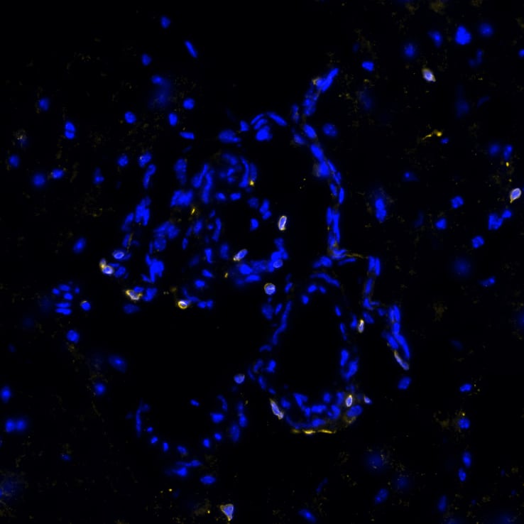
Detection of STING/TMEM173 in Human Brain Cortex (Cerebrum) via seqIF™ staining on COMET™
STING/TMEM173 was detected in immersion fixed paraffin-embedded sections of human brain Cortex (Cerebrum) using Rabbit Anti-Human STING/TMEM173, Monoclonal Antibody (Catalog #NBP3-18816) at 10ug/mL at 37 ° Celsius for 4 minutes. Before incubation with the primary antibody, tissue underwent an all-in-one dewaxing and antigen retrieval preprocessing using PreTreatment Module (PT Module) and Dewax and HIER Buffer H (pH 9; Epredia Catalog # TA-999-DHBH). Tissue was stained using the Alexa Fluor™ Plus 647 Goat anti-Rabbit IgG Secondary Antibody at 1:200 at 37 ° Celsius for 2 minutes. (Yellow; Lunaphore Catalog # DR647RB) and counterstained with DAPI (blue; Lunaphore Catalog # DR100). Specific staining was localized to the membrane. Protocol available in COMET™ Panel Builder.The image is attached.![STING/TMEM173 Antibody (2922D) - BSA Free Multiplex Immunofluorescence: STING/TMEM173 Antibody (2922D) - BSA Free [NBP3-18816] -](https://resources.bio-techne.com/images/products/nbp3-18816_-multiplex-immunofluorescence-639186281958742538.png "Detection of STING in Frozen Mouse Hippocampus via seqIF™ staining on COMET™")
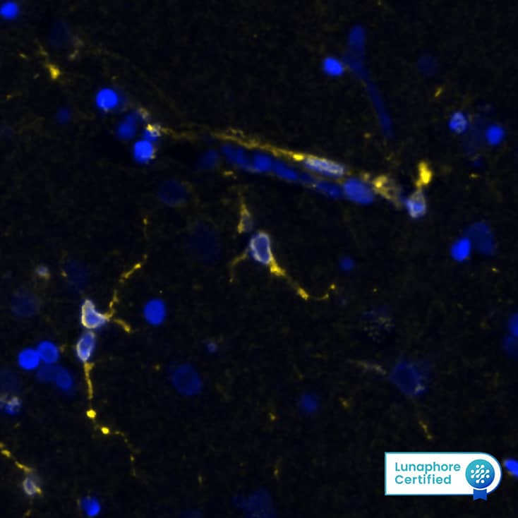
Detection of STING in Frozen Mouse Hippocampus via seqIF™ staining on COMET™
STING was detected in frozen sections of mouse hippocampus using Rabbit Anti-Mouse STING Monoclonal Antibody (Catalog # NBP3-18816) at 10ug/mL at 37 ° Celsius for 4 minutes. Before incubation with the primary antibody, tissue underwent preprocessing by incubating tissue with Multi Staining Buffer (Lunaphore Catalog # BU06) for 5minutes at room temperature followed by a 20-minute incubation in Tris-Buffered Saline + 0.2% Triton at room temperature. Tissue was stained using the Alexa Fluor™ Plus 647 Goat anti-Rabbit IgG Secondary Antibody at 1:200 at 37 ° Celsius for 2 minutes. (Yellow; Lunaphore Catalog # DR647RB) and counterstained with DAPI (blue; Lunaphore Catalog # DR100). Specific staining was localized to the cytoplasm. Protocol available in COMET™ Panel Builder.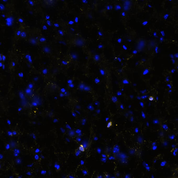
Detection of STING in Frozen Mouse Cerebral Cortex via seqIF™ staining on COMET™
STING was detected in frozen sections of mouse cerebral cortex using Rabbit Anti-Mouse STING Monoclonal Antibody (Catalog # NBP3-18816) at 10ug/mL at 37 ° Celsius for 4 minutes. Before incubation with the primary antibody, tissue underwent preprocessing by incubating tissue with Multi Staining Buffer (Lunaphore Catalog # BU06) for 5minutes at room temperature followed by a 20-minute incubation in Tris-Buffered Saline + 0.2% Triton at room temperature. Tissue was stained using the Alexa Fluor™ Plus 647 Goat anti-Rabbit IgG Secondary Antibody at 1:200 at 37 ° Celsius for 2 minutes. (Yellow; Lunaphore Catalog # DR647RB) and counterstained with DAPI (blue; Lunaphore Catalog # DR100). Specific staining was localized to the cytoplasm. Protocol available in COMET™ Panel Builder.![Western Blot: STING/TMEM173 Antibody (2922D) [NBP3-18816]](https://resources.bio-techne.com/images/products/STING-TMEM173-Antibody-2922D-Western-Blot-NBP3-18816-img0003.jpg "Western Blot: STING/TMEM173 Antibody (2922D) [NBP3-18816]")
![Immunocytochemistry/ Immunofluorescence: STING/TMEM173 Antibody (2922D) [NBP3-18816]](https://resources.bio-techne.com/images/products/STING-TMEM173-Antibody-2292D-Immunocytochemistry-Immunofluorescence-NBP3-18816-img0002.jpg "Immunocytochemistry/ Immunofluorescence: STING/TMEM173 Antibody (2922D) [NBP3-18816]")
![Immunohistochemistry: STING/TMEM173 Antibody (2922D) [NBP3-18816]](https://resources.bio-techne.com/images/products/STING-TMEM173-Antibody-2292D-Immunohistochemistry-NBP3-18816-img0001.jpg "Immunohistochemistry: STING/TMEM173 Antibody (2922D) [NBP3-18816]")
![Simple Western: STING/TMEM173 Antibody (2922D) [NBP3-18816]](https://resources.bio-techne.com/images/products/STING-TMEM173-Antibody-2922D-Simple-Western-NBP3-18816-img0004.jpg "Simple Western: STING/TMEM173 Antibody (2922D) [NBP3-18816]")


Applications for STING/TMEM173 Antibody (2922D) - BSA Free
Application
Recommended Usage
Immunocytochemistry/ Immunofluorescence
3 ug/ml
Immunohistochemistry
0.5 ug/ml
Immunoprecipitation
Validated for Immunoprecipitation from YCharOS Inc. (ycharos.com)
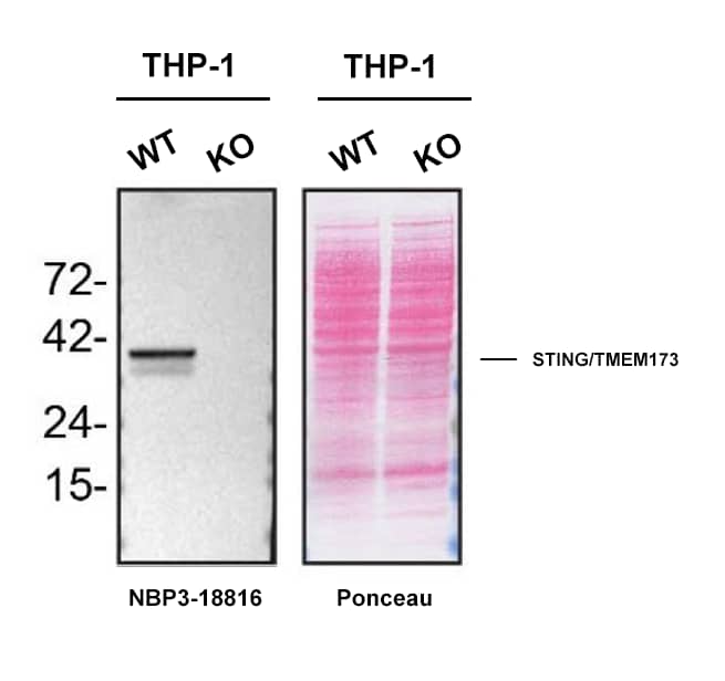
Knockout Validated
Validated for Knockout from YCharOS Inc. (ycharos.com)
Multiplex Immunofluorescence
10 ug/mL
Western Blot
1 - 2 ug/ml
Formulation, Preparation, and Storage
Purification
Protein A or G purified
Formulation
PBS
Format
BSA Free
Preservative
0.02% Sodium Azide
Concentration
1.0 mg/ml
Shipping
The product is shipped with polar packs. Upon receipt, store it immediately at the temperature recommended below.
Stability & Storage
Store at 4C for up to 3 months. For longer storage, aliquot and store at -20C.
Background: STING/TMEM173
References
1. Patel, S., & Jin, L. (2019). TMEM173 variants and potential importance to human biology and disease. Genes and Immunity. https://doi.org/10.1038/s41435-018-0029-9
2. Jounai, N., Kobiyama, K., Takeshita, F., & Ishii, K. J. (2013). Recognition of damage-associated molecular patterns related to nucleic acids during inflammation and vaccination. Frontiers in Cellular and Infection Microbiology. https://doi.org/10.3389/fcimb.2012.00168
3. Xiao, T. S., & Fitzgerald, K. A. (2013). The cGAS-STING Pathway for DNA Sensing. Molecular Cell. https://doi.org/10.1016/j.molcel.2013.07.004
4. Kato, K., Omura, H., Ishitani, R., & Nureki, O. (2017). Cyclic GMP-AMP as an Endogenous Second Messenger in Innate Immune Signaling by Cytosolic DNA. Annual Review of Biochemistry. https://doi.org/10.1146/annurev-biochem-061516-044813
5. Crowl, J. T., Gray, E. E., Pestal, K., Volkman, H. E., & Stetson, D. B. (2017). Intracellular Nucleic Acid Detection in Autoimmunity. Annual Review of Immunology. https://doi.org/10.1146/annurev-immunol-051116-052331
Long Name
Stimulator of Interferon Genes Protein/Transmembrane protein 173
Alternate Names
ERIS, MITA, MPYS, NET23, TMEM173
Gene Symbol
STING1
Additional STING/TMEM173 Products
Product Documents for STING/TMEM173 Antibody (2922D) - BSA Free
Product Specific Notices for STING/TMEM173 Antibody (2922D) - BSA Free
This product is for research use only and is not approved for use in humans or in clinical diagnosis. Primary Antibodies are guaranteed for 1 year from date of receipt.
Loading...
Loading...
Loading...
Loading...