
Human/Mouse Ki67/MKI67 Antibody
R&D Systems, part of Bio-Techne | Catalog # MAB7617
Recombinant Monoclonal Antibody.

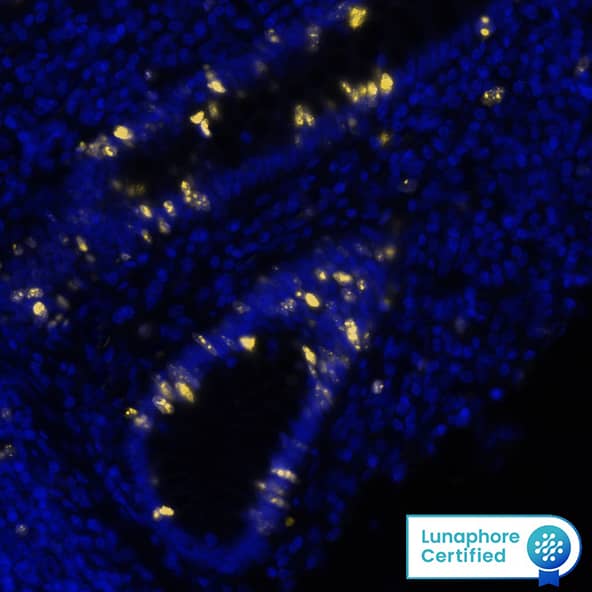
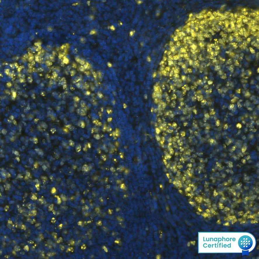
Scientific images may be resized, cropped, or adjusted for brightness or contrast for presentation purposes. These updates are limited to presentation and do not affect the underlying scientific interpretation of the data. Where available, additional source data may be provided upon request.
Conjugate
Catalog #
Key Product Details
Validated by
Knockout/Knockdown
Species Reactivity
Validated:
Human, Mouse
Cited:
Human, Mouse
Applications
Validated:
Knockout Validated, Immunohistochemistry, Intracellular Staining by Flow Cytometry, Dual RNAscope ISH-IHC Compatible, Immunocytochemistry, Simple Western, COMET
Cited:
Immunohistochemistry, Immunocytochemistry, Immunocytochemistry/ Immunofluorescence
Label
Unconjugated
Antibody Source
Recombinant Monoclonal Rabbit IgG Clone # 1297A
Product Specifications
Immunogen
Mouse Ki67/MKI67 synthetic peptide
Accession # E9PVX6
Accession # E9PVX6
Specificity
Detects human Ki67/MKI67 in direct ELISAs.
Clonality
Monoclonal
Host
Rabbit
Isotype
IgG
Scientific Data Images for Human/Mouse Ki67/MKI67 Antibody
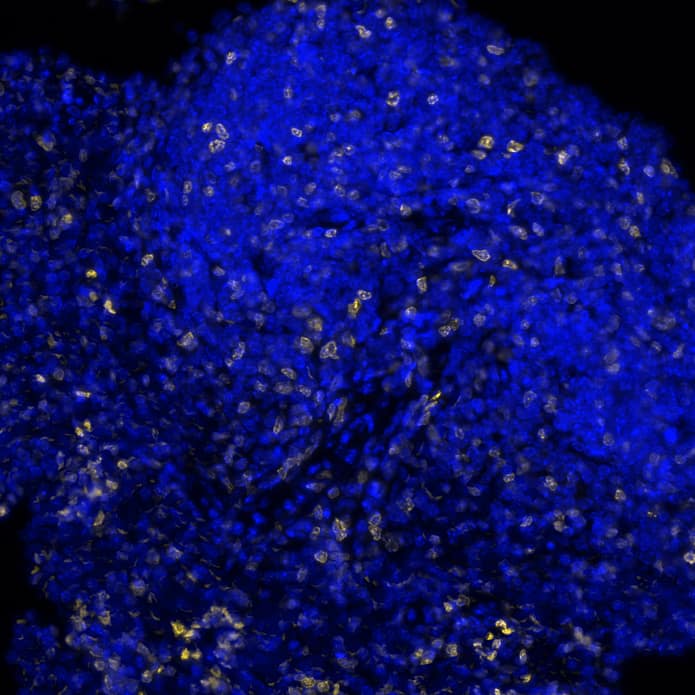
Detection of Ki67/MKI67 in Human Tonsil via seqIF™ staining on COMET™
Ki67/MKI67 was detected in immersion fixed paraffin-embedded sections of human tonsil using Rabbit Anti-Human Ki67/MKI67 Monoclonal Antibody (Catalog # MAB7617) at 10 µg/mL at 37 ° Celsius for 4 minutes. Before incubation with the primary antibody, tissue underwent an all-in-one dewaxing and antigen retrieval preprocessing using PreTreatment Module (PT Module) and Dewax and HIER Buffer H (pH 9). Tissue was stained using the Alexa Fluor™ Plus 555 Goat anti-Rabbit IgG Secondary Antibody at 1:100 at 37 ° Celsius for 2 minutes. (Yellow; Lunaphore Catalog # DR555RB) and counterstained with DAPI (blue; Lunaphore Catalog # DR100).. Specific staining was localized to the nucleus. Protocol available in COMET™ Panel Builder.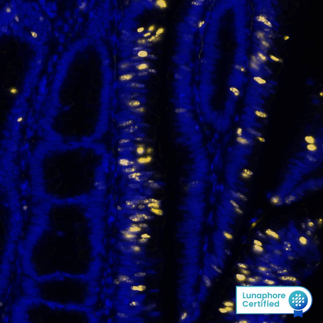
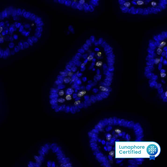
Detection of Ki67/MKI67 in Human Colon via seqIF™ staining on COMET™
Ki67/MKI67 was detected in immersion fixed paraffin-embedded sections of human Colon using Rabbit Anti-Human Ki67/MKI67 Monoclonal Antibody (Catalog # MAB7617) at 5 µg/mL at 37° Celsius for 4 minutes. Before incubation with the primary antibody, tissue underwent an all-in-one dewaxing and antigen retrieval preprocessing using PreTreatment Module (PT Module) and Dewax and HIER Buffer H (pH 9). Tissue was stained using the Alexa Fluor™ Plus 555 Goat anti-Rabbit IgG Secondary Antibody at 1:100 at 37° Celsius for 2 minutes. (Yellow; Lunaphore Catalog # DR555RB) and counterstained with DAPI (blue; Lunaphore Catalog # DR100).. Specific staining was localized to the nucleus. Protocol available in COMET™ Panel Builder.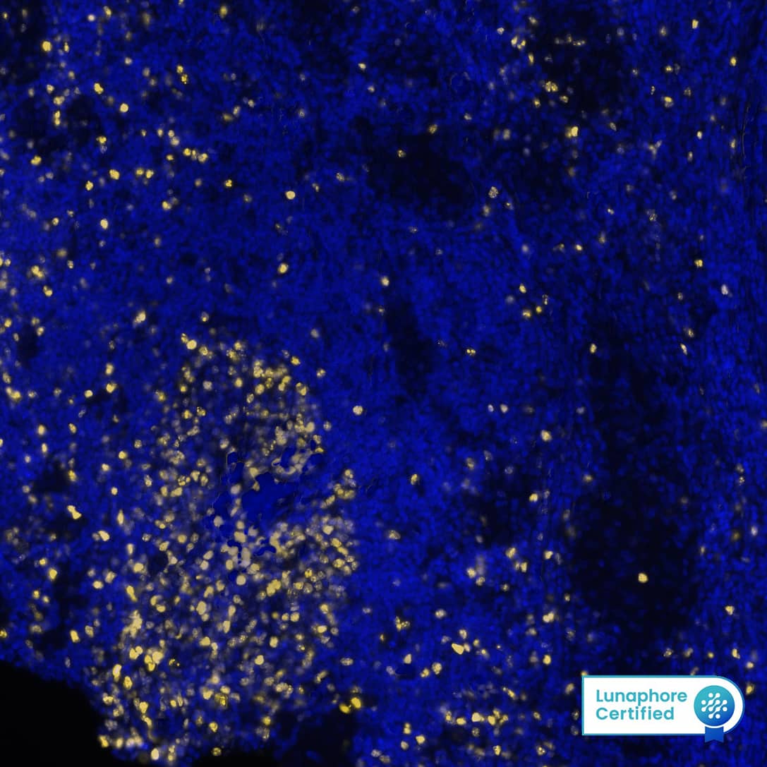
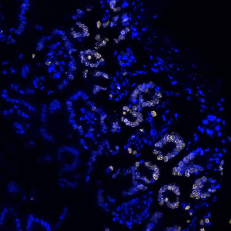
Detection of Ki67/MKI67 in Human Hodgkin Lymphoma via seqIF™ staining on COMET™
Ki67/MKI67 was detected in immersion fixed paraffin-embedded sections of human Hodgkin Lymphoma using Rabbit Anti-Human Ki67/MKI67 Monoclonal Antibody (Catalog # MAB7617) at 5 µg/mL at 37 ° Celsius for 4 minutes. Before incubation with the primary antibody, tissue underwent an all-in-one dewaxing and antigen retrieval preprocessing using PreTreatment Module (PT Module) and Dewax and HIER Buffer H (pH 9). Tissue was stained using the Alexa Fluor™ Plus 555 Goat anti-Rabbit IgG Secondary Antibody at 1:100 at 37 ° Celsius for 2 minutes. (Yellow; Lunaphore Catalog # DR555RB) and counterstained with DAPI (blue; Lunaphore Catalog # DR100).. Specific staining was localized to the nucleus. Protocol available in COMET™ Panel Builder.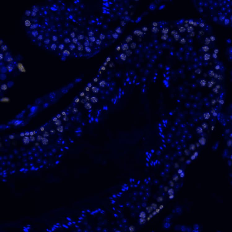
Applications for Human/Mouse Ki67/MKI67 Antibody
Application
Recommended Usage
COMET
0.15-10 µg/mL
Sample: Immersion fixed paraffin-embedded sections of human tonsil, colon, Hodgkin Lymphoma and appendix, and mouse intestine, stomach, testis and spleen
Sample: Immersion fixed paraffin-embedded sections of human tonsil, colon, Hodgkin Lymphoma and appendix, and mouse intestine, stomach, testis and spleen
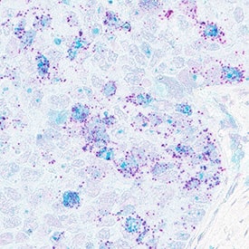
Dual RNAscope ISH-IHC Compatible
5-25 µg/mL
Sample: Formalin-fixed paraffin-embedded tissue sections of human breast cancer
Sample: Formalin-fixed paraffin-embedded tissue sections of human breast cancer
Immunocytochemistry
0.3-25 µg/mL
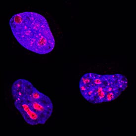
Sample: Immersion fixed HeLa human cervical epithelial carcinoma cell line
Sample: Immersion fixed HeLa human cervical epithelial carcinoma cell line
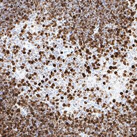
Immunohistochemistry
3-25 µg/mL
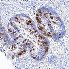
Sample: Immersion fixed paraffin-embedded sections of human pancreatic cancer tissue and mouse thymus
Sample: Immersion fixed paraffin-embedded sections of human pancreatic cancer tissue and mouse thymus
Intracellular Staining by Flow Cytometry
0.25 µg/106 cells
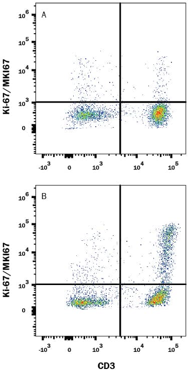
Sample: Human peripheral blood mononuclear cells (PBMCs) treated with PHA were fixed and permeabilized with FlowX FoxP3 Fixation & Permeabilization Buffer Kit (Catalog # FC012)
Sample: Human peripheral blood mononuclear cells (PBMCs) treated with PHA were fixed and permeabilized with FlowX FoxP3 Fixation & Permeabilization Buffer Kit (Catalog # FC012)
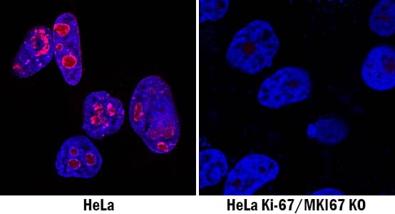
Knockout Validated
Ki67/MKI67 is specifically detected in Hela human cervical epithelial carcinoma parental cell line but is not detectable in Ki67/MKI67 knockout HeLa cell line.
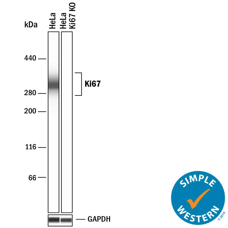
Simple Western
20 µg/mL
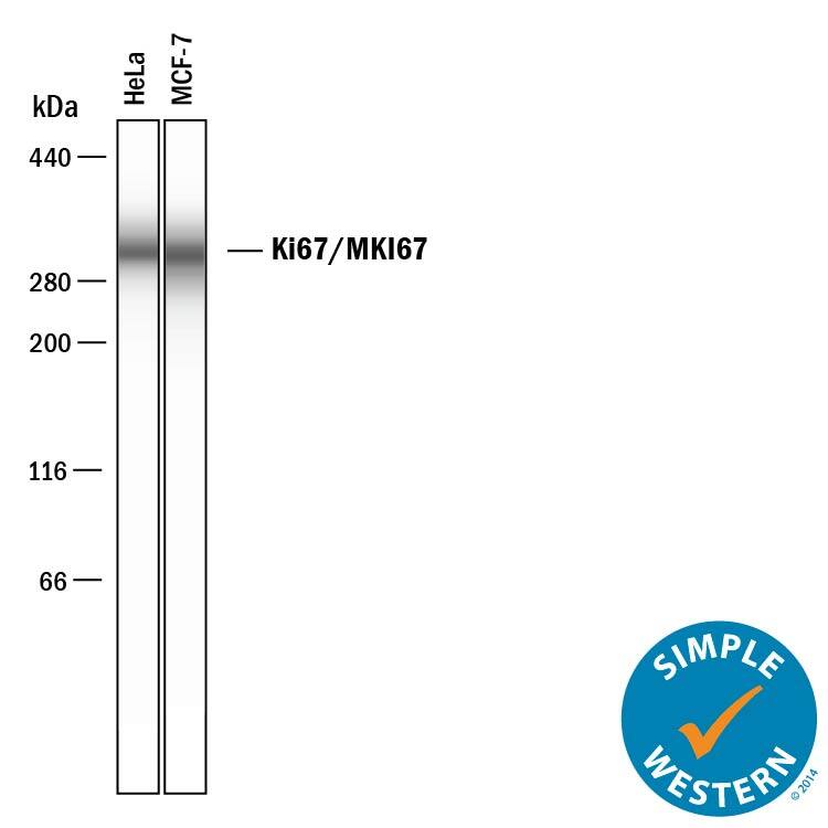
Sample: HeLa human cervical epithelial carcinoma cell line and MCF‑7 human breast cancer cell line
Sample: HeLa human cervical epithelial carcinoma cell line and MCF‑7 human breast cancer cell line
Reviewed Applications
Read 3 reviews rated 4.3 using MAB7617 in the following applications:
Formulation, Preparation, and Storage
Purification
Protein A or G purified from cell culture supernatant
Reconstitution
Reconstitute at 0.5 mg/mL in sterile PBS. For liquid material, refer to CoA for concentration.
Formulation
Lyophilized from a 0.2 μm filtered solution in PBS with Trehalose. See Certificate of Analysis for details.
*Small pack size (-SP) is supplied either lyophilized or as a 0.2 µm filtered solution in PBS.
*Small pack size (-SP) is supplied either lyophilized or as a 0.2 µm filtered solution in PBS.
Shipping
Lyophilized product is shipped at ambient temperature. Liquid small pack size (-SP) is shipped with polar packs. Upon receipt, store immediately at the temperature recommended below.
Stability & Storage
Use a manual defrost freezer and avoid repeated freeze-thaw cycles.
- 12 months from date of receipt, -20 to -70 °C as supplied.
- 1 month, 2 to 8 °C under sterile conditions after reconstitution.
- 6 months, -20 to -70 °C under sterile conditions after reconstitution.
Background: Ki67/MKI67
MKI67 (also Ki67) is a 350-400 kDa nuclear protein that belongs to a molecular group comprised of mitotic chromosome-associated proteins. Ki67 was originally recognized as an antigen associated with the monoclonal Ki67 antibody raised against Hodgkin's lymphoma nuclear material. Ki-67 is contextually expressed, being potentially found in all cells that are not in the Go phase of the cell cycle. Thus, MKI67 qualifies as a cell proliferation marker. Functionally, Ki67 is known to interact with 160 kDa Hklp2, a protein that promotes centrosome separation and spindle bipolarity. It also directly interacts with NIFK, and apparently binds to UBF, thus playing a role in rRNA synthesis. Human MKI67 is 3256 amino acids (aa) in length. It contains one FHA domain (aa 8-98), followed by at least 24 utilized Ser/Thr phosphorylation sites and sixteen 120 aa repeats (aa 1000-2928) that are interspersed with at least 90 additional utilized phosphorylation sites. There are two potential isoform variants. One isoform is 315-345 kDa in size and shows a deletion of aa 136-495, while a second isoform contains a 58 aa substitution for aa 1-513. Over aa 3120-3256, human Ki67 shares 46% aa sequence identity with the mouse ortholog to Ki67.
Long Name
Antigen Identified by Monoclonal Antibody Ki67
Alternate Names
Ki-67, KIA, MIB-1, MKI67, PPP1R105, TSG126
Gene Symbol
MKI67
UniProt
Additional Ki67/MKI67 Products
Product Documents for Human/Mouse Ki67/MKI67 Antibody
Product Specific Notices for Human/Mouse Ki67/MKI67 Antibody
For research use only
Loading...
Loading...
Loading...
Loading...
Loading...
Loading...