
Human IL-13 R alpha2 Antibody
R&D Systems, part of Bio-Techne | Catalog # AF146

Scientific images may be resized, cropped, or adjusted for brightness or contrast for presentation purposes. These updates are limited to presentation and do not affect the underlying scientific interpretation of the data. Where available, additional source data may be provided upon request.
Conjugate
Catalog #
Key Product Details
Validated by
Knockout/Knockdown, Biological Validation
Species Reactivity
Validated:
Human
Cited:
Human, Mouse, Xenograft
Applications
Validated:
Immunohistochemistry, Western Blot, Blockade of Receptor-ligand Interaction, Flow Cytometry, Simple Western, CyTOF-ready
Cited:
Immunohistochemistry, Immunohistochemistry-Paraffin, Western Blot, Neutralization, Flow Cytometry, Immunocytochemistry, Immunoprecipitation, CAR-T (Flow Cytometry), CAR-T (IHC-P)
Label
Unconjugated
Antibody Source
Polyclonal Goat IgG
Product Specifications
Immunogen
Mouse myeloma cell line NS0-derived recombinant human IL-13 R alpha2
Cys22-Leu342
Accession # Q14627
Cys22-Leu342
Accession # Q14627
Specificity
Detects human IL-13 R alpha2 in direct ELISAs. In direct ELISAs, approximately 5% cross-reactivity with recombinant mouse IL-13 R alpha2 is observed and less than 1% cross-reactivity with recombinant human IL-13 R alpha1 is observed.
Clonality
Polyclonal
Host
Goat
Isotype
IgG
Endotoxin Level
<0.10 EU per 1 μg of the antibody by the LAL method.
Scientific Data Images for Human IL-13 R alpha2 Antibody
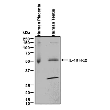
Detection of Human IL‑13 R alpha2 by Western Blot.
Western blot shows lysates of human placenta tissue and human testis tissue. PVDF membrane was probed with 0.2 µg/mL of Goat Anti-Human IL-13 Ra2 Antigen Affinity-purified Polyclonal Antibody (Catalog # AF146) followed by HRP-conjugated Anti-Goat IgG Secondary Antibody HAF017). A specific band was detected for IL-13 Ra2 at approximately 50-55 kDa (as indicated). This experiment was conducted under reducing conditions and using Immunoblot Buffer Group 1.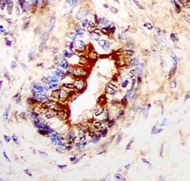
IL-13 R alpha2 in Human Prostate Cancer Tissue.
IL-13 Ra2 was detected in immersion fixed paraffin-embedded sections of human prostate cancer tissue using Goat Anti-Human IL-13 Ra2 Antigen Affinity-purified Polyclonal Antibody (Catalog # AF146) at 5 µg/mL overnight at 4 °C. Tissue was stained using the Anti-Goat HRP-DAB Cell & Tissue Staining Kit (brown CTS008) and counterstained with hematoxylin (blue). Specific staining was localized to glandular epithelial cells. View our protocol for Chromogenic IHC Staining of Paraffin-embedded Tissue Sections.
Detection of Human IL‑13 R alpha2 by Simple WesternTM.
Simple Western lane view shows lysates of human placenta tissue and human testis tissue, loaded at 0.2 mg/mL. A specific band was detected for IL-13 Ra2 at approximately 59 kDa (as indicated) using 10 µg/mL of Goat Anti-Human IL-13 Ra2 Antigen Affinity-purified Polyclonal Antibody (Catalog # AF146) followed by 1:50 dilution of HRP-conjugated Anti-Goat IgG Secondary Antibody (Catalog # HAF109). This experiment was conducted under reducing conditions and using the 12-230 kDa separation system.







Applications for Human IL-13 R alpha2 Antibody
Application
Recommended Usage
Blockade of Receptor-ligand Interaction
In a functional ELISA, 2-6 µg/mL of this antibody will block 50% of the binding of 100 ng/mL of Recombinant Human IL-13 (Catalog # 213‑ILB) to immobilized Recombinant Human IL-13 R alpha2 Fc Chimera (Catalog # 614-INS) coated at 4 µg/mL (100 µL/well). At 40 μg/mL, this antibody will block >90% of the binding.
CyTOF-ready
Ready to be labeled using established conjugation methods. No BSA or other carrier proteins that could interfere with conjugation.
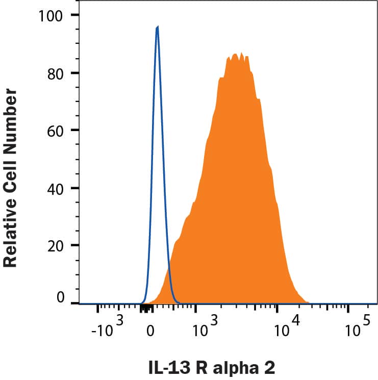
Flow Cytometry
2.5 µg/106 cells
Sample: A375 human melanoma cell line
Sample: A375 human melanoma cell line
Immunohistochemistry
5-15 µg/mL
Sample: Immersion fixed paraffin-embedded sections of human normal skin and prostate cancer tissue
Sample: Immersion fixed paraffin-embedded sections of human normal skin and prostate cancer tissue
Simple Western
10 µg/mL
Sample: Human placenta tissue and human testis tissue
Sample: Human placenta tissue and human testis tissue
Western Blot
0.2 µg/mL
Sample: Human placenta tissue and human testis tissue
Sample: Human placenta tissue and human testis tissue
Formulation, Preparation, and Storage
Purification
Antigen Affinity-purified
Reconstitution
Reconstitute at 0.2 mg/mL in sterile PBS. For liquid material, refer to CoA for concentration.
Formulation
Lyophilized from a 0.2 μm filtered solution in PBS with Trehalose. See Certificate of Analysis for details.
*Small pack size (-SP) is supplied either lyophilized or as a 0.2 µm filtered solution in PBS.
*Small pack size (-SP) is supplied either lyophilized or as a 0.2 µm filtered solution in PBS.
Shipping
Lyophilized product is shipped at ambient temperature. Liquid small pack size (-SP) is shipped with polar packs. Upon receipt, store immediately at the temperature recommended below.
Stability & Storage
Use a manual defrost freezer and avoid repeated freeze-thaw cycles.
- 12 months from date of receipt, -20 to -70 °C as supplied.
- 1 month, 2 to 8 °C under sterile conditions after reconstitution.
- 6 months, -20 to -70 °C under sterile conditions after reconstitution.
Background: IL-13 R alpha 2
Two type1 membrane proteins belonging to the hemopoietin receptor family have been cloned and shown to bind IL-13 with differing affinities. The lower affinity IL-13 binding protein, previously designated IL-13 R alpha, IL-13 R alphal or NR4, is now referred to as IL-13 R alpha1. The high affinity IL-13 binding protein, previously also designated IL-13 R or IL-13 R alphal, is now referred to as IL-13 R alpha2. Human IL-13 R alpha2 was originally cloned from the Caki-1 human renal carcinoma cell line. The IL-13 R alpha2 cDNA encodes a 380 amino acid (aa) residue precursor protein with a putative 26 aa residue signal peptide, a 317 residue extracellular domain, a 20 aa residue transmembrane region and a 17 aa residue cytoplasmic tail. Human and mouse IL‑13 R alpha2 share 59% aa sequence identity. The extracellular domain of IL-13 R alpha2 is also closely related to that of IL-13 R alpha1. However, the 17 aa residue cytoplasmic domain of IL-13 R alpha2 is much shorter than that of IL-13 R alpha1, suggesting that the two receptors are functionally distinct. IL-13 R alpha1 has been shown to combine with the IL-4 R to form a high-affinity receptor complex capable of transducing an IL‑13‑dependent proliferative signal. The role of IL-13 R alpha2 in IL-13 signaling remains to be elucidated. The amino-terminal 27 aa residues of the human and mouse IL‑13 R alpha2 are nearly identical to that of a soluble mouse IL-13 binding protein purified from mouse serum and urine.
References
- Caput, D. et al. (1996) J. Biol. Chem. 271:16921.
- Donaldson, D.D. et al. (1998) J. Immunol. 161:2317.
- Aman, M.J. et al. (1996) J. Biol. Chem. 271:29265.
- Hilton, D.J. et al. (1996) Proc. Natl. Acad. Sci. USA 93:497.
- Zhang, J.G. et al. (1997) J. Biol. Chem. 272:9474.
Long Name
Interleukin 13 Receptor alpha 2
Alternate Names
CD213a2, IL-13Ra2, IL13R alpha 2, IL13RA2
Gene Symbol
IL13RA2
UniProt
Additional IL-13 R alpha 2 Products
Product Documents for Human IL-13 R alpha2 Antibody
Product Specific Notices for Human IL-13 R alpha2 Antibody
For research use only
Loading...
Loading...
Loading...
Loading...
Loading...
Loading...