
Human FUS Antibody
R&D Systems, part of Bio-Techne | Catalog # MAB11793

Conjugate
Catalog #
Key Product Details
Species Reactivity
Human
Applications
Immunohistochemistry, Western Blot, Simple Western
Label
Unconjugated
Antibody Source
Monoclonal Mouse IgG2B Clone # 1120264)
Product Specifications
Immunogen
E. coli - derived recombinant human FUS
Asn263-Gly378
Accession # P35637
Asn263-Gly378
Accession # P35637
Specificity
Detects recombinant human FUS and human TAF-15 in Direct ELISA.
Clonality
Monoclonal
Host
Mouse
Isotype
IgG2B
Scientific Data Images for Human FUS Antibody
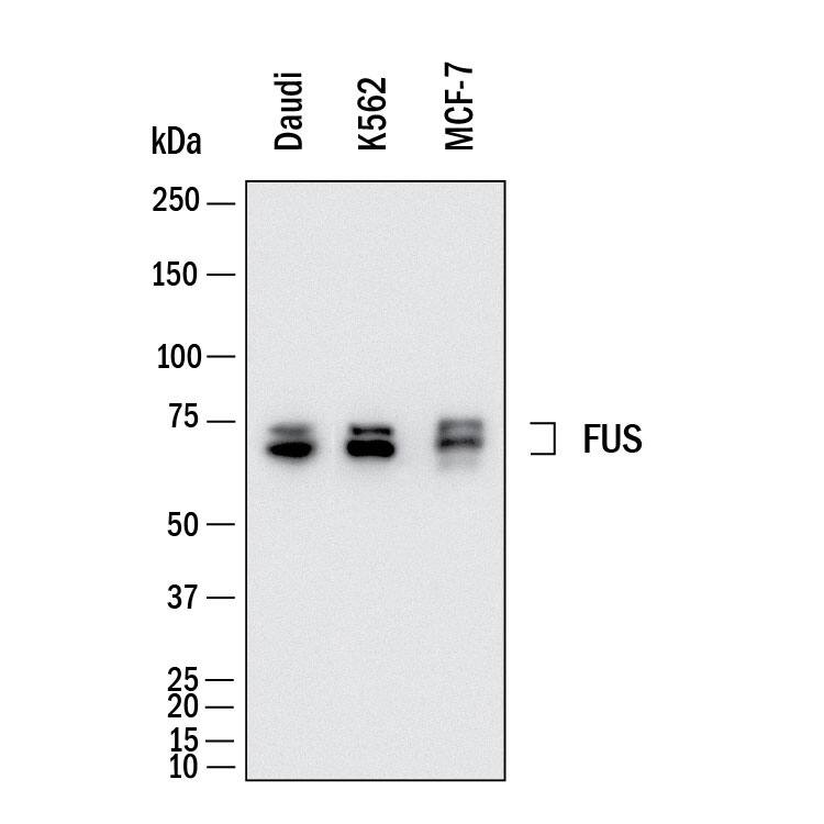
Detection of Human FUS by Western Blot.
Western Blot shows lysates of Daudi human Burkitt's lymphoma cell line, K562 human chronic myelogenous leukemia cell line and MCF-7 human breast cancer cell line. PVDF membrane was probed with 0.5 µg/ml of Mouse Anti-Human FUS Monoclonal Antibody (Catalog # MAB11793) followed by HRP-conjugated Anti-Mouse IgG Secondary Antibody (HAF018). Specific bands were detected for FUS at approximately 70 kDa (as indicated). This experiment was conducted under reducing conditions and using Western Blot Buffer Group 1.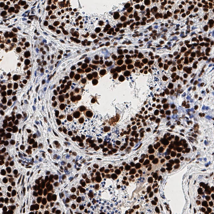
Immersion fixed paraffin-embedded sections of human testis
FUS was detected in immersion fixed paraffin-embedded sections of human testis using Mouse Anti-Human FUS Monoclonal Antibody (Catalog # MAB11793) at 5 µg/ml overnight at 4 °C. Before incubation with the primary antibody, tissue was subjected to heat-induced epitope retrieval using VisUCyte Antigen Retrieval Reagent-Basic (VCTS021). Tissue was stained using the HRP-conjugated Anti-Mouse IgG Secondary Antibody (HAF007) and counterstained with hematoxylin (blue). Specific staining was localized to the nucleus. View our protocol for Chromogenic IHC Staining of Paraffin-embedded Tissue Sections.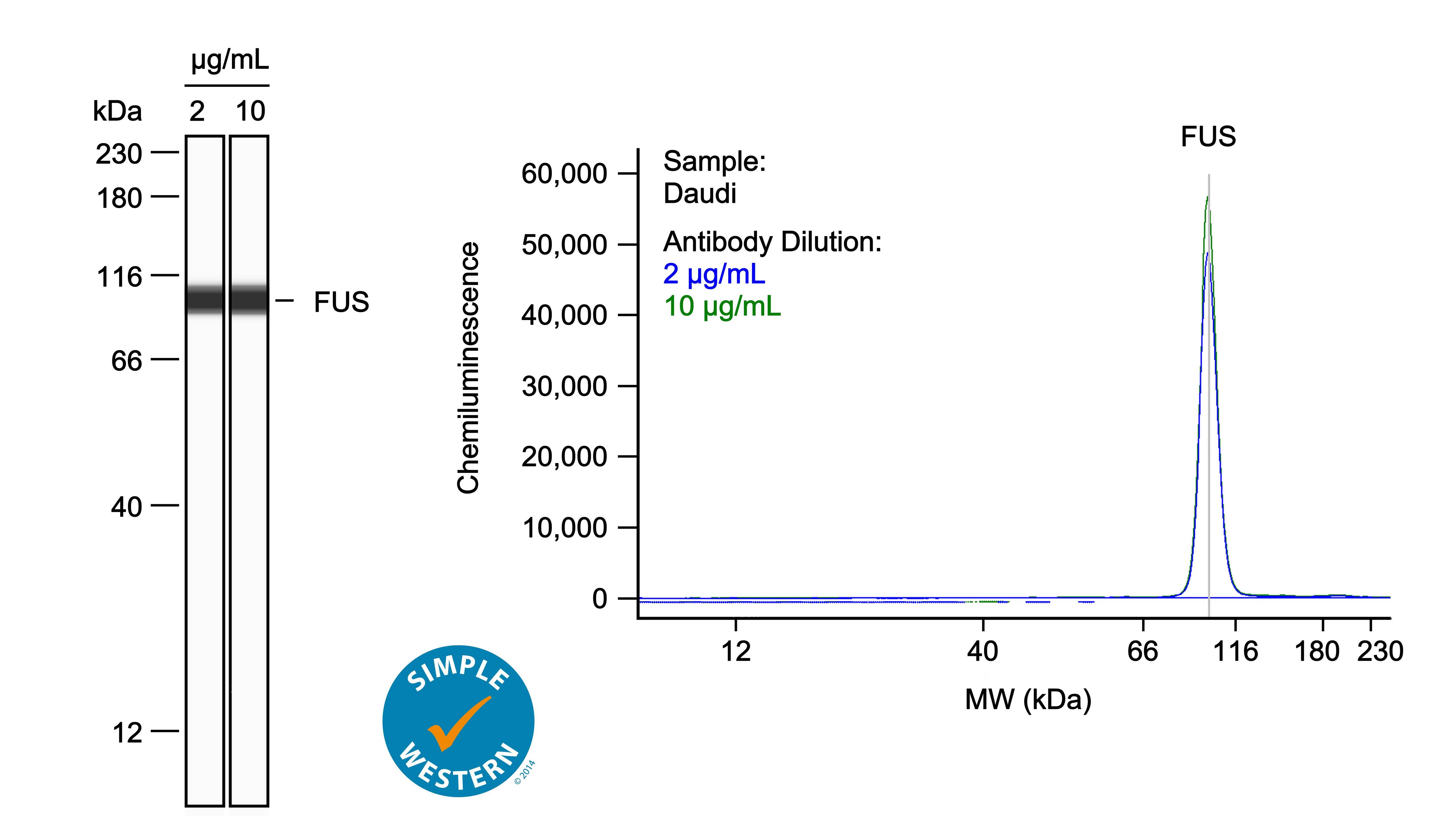
Detection of Human FUS by Simple WesternTM.
Left: Simple Western lane view shows lysates of Daudi human Burkitt's lymphoma cell line, loaded at 0.1 mg/ml. A specific band was detected for FUS at approximately 101 kDa (as indicated) using both 2 µg/ml and 10 µg/ml of Mouse Anti-Human FUS Monoclonal Antibody (Catalog # MAB11793) followed by HRP-conjugated Goat Anti-Mouse Secondary Antibody (042-205). This experiment was conducted under reducing conditions and using the 12-230kDa separation system. Right: Simple Western electropherogram showing the same Mouse Anti-Human FUS Monoclonal Antibody (Catalog # MAB11793) tested at 2 µg/ml (blue line) and 10 µg/ml (green line) in the Daudi human Burkitt's lymphoma cell line.Applications for Human FUS Antibody
Application
Recommended Usage
Immunohistochemistry
0.25-25 µg/mL
Sample: Immersion fixed paraffin-embedded sections of human testis
Sample: Immersion fixed paraffin-embedded sections of human testis
Simple Western
2-10 µg/mL
Sample: Daudi human Burkitt's lymphoma cell line
Sample: Daudi human Burkitt's lymphoma cell line
Western Blot
0.5 µg/mL
Sample: Daudi human Burkitt's lymphoma cell line, K562 human chronic myelogenous leukemia cell line and MCF-7 human breast cancer cell line
Sample: Daudi human Burkitt's lymphoma cell line, K562 human chronic myelogenous leukemia cell line and MCF-7 human breast cancer cell line
Formulation, Preparation, and Storage
Purification
Protein A or G purified from hybridoma culture supernatant
Reconstitution
Reconstitute lyophilized material at 0.2 mg/ml in sterile PBS. For liquid material, refer to CoA for concentration.
Formulation
Lyophilized from a 0.2 μm filtered solution in PBS with Trehalose.
Shipping
Lyophilized product is shipped at ambient temperature. Liquid small pack size (-SP) is shipped with polar packs. Upon receipt, store immediately at the temperature recommended below.
Stability & Storage
Use a manual defrost freezer and avoid repeated freeze-thaw cycles.
- 12 months from date of receipt, -20 to -70 °C as supplied.
- 1 month, 2 to 8 °C under sterile conditions after reconstitution.
- 6 months, -20 to -70 °C under sterile conditions after reconstitution.
Background: FUS
References
- Vance C, Rogelj B, Hortobágyi T, De Vos KJ, Nishimura AL, Sreedharan J, Hu X, Smith B, Ruddy D, Wright P, Ganesalingam J, Williams KL, Tripathi V, Al-Saraj S, Al-Chalabi A, Leigh PN, Blair IP, Nicholson G, de Belleroche J, Gallo JM, Miller CC, Shaw CE. Mutations in FUS, an RNA processing protein, cause familial amyotrophic lateral sclerosis type 6. Science. 2009 Feb 27;323(5918):1208-1211. doi: 10.1126/science.1165942. PMID: 19251628; PMCID: PMC4516382.
- Sama RR, Ward CL, Bosco DA. Functions of FUS/TLS from DNA repair to stress response: implications for ALS. ASN Neuro. 2014 Jun 1;6(4):1759091414544472. doi: 10.1177/1759091414544472. PMID: 25289647; PMCID: PMC4189536.
- Yang S, Warraich ST, Nicholson GA, Blair IP. Fused in sarcoma/translocated in liposarcoma: a multifunctional DNA/RNA binding protein. Int J Biochem Cell Biol. 2010 Sep;42(9):1408-11. doi: 10.1016/j.biocel.2010.06.003. Epub 2010 Jun 10. PMID: 20541619.
Long Name
FUS RNA binding protein
Alternate Names
ALS6, altFUS, ETM4, FUS1, HNRNPP2, POMP75, TLS
Gene Symbol
FUS
UniProt
Additional FUS Products
Product Documents for Human FUS Antibody
Product Specific Notices for Human FUS Antibody
For research use only
Loading...
Loading...
Loading...
Loading...
Loading...