
Equine IL-10 Antibody
R&D Systems, part of Bio-Techne | Catalog # AF1605



Scientific images may be resized, cropped, or adjusted for brightness or contrast for presentation purposes. These updates are limited to presentation and do not affect the underlying scientific interpretation of the data. Where available, additional source data may be provided upon request.
Conjugate
Catalog #
Key Product Details
Validated by
Biological Validation
Species Reactivity
Validated:
Equine
Cited:
Mouse
Applications
Validated:
Western Blot, ELISA Capture (Matched Antibody Pair), Neutralization, Immunocytochemistry
Cited:
Immunohistochemistry-Frozen
Label
Unconjugated
Antibody Source
Polyclonal Goat IgG
Product Specifications
Immunogen
E. coli-derived recombinant equine IL-10
Ser19-Asn178
Accession # Q28374
Ser19-Asn178
Accession # Q28374
Specificity
Detects equine IL‑10 in ELISAs and Western blots. In sandwich immunoassays, less than 1% cross-reactivity with recombinant canine IL-10 and recombinant porcine IL-10 is observed and less than 0.4% cross-reactivity with recombinant human IL-10, recombinant mouse IL-10, recombinant rat IL-10, and recombinant feline IL-10 is observed.
Clonality
Polyclonal
Host
Goat
Isotype
IgG
Endotoxin Level
<0.15 EU per 1 μg of the antibody by the LAL method.
Scientific Data Images for Equine IL-10 Antibody
Cell Proliferation Induced by IL‑10 and Neutralization by Equine IL‑10 Antibody.
Recombinant Equine IL-10 (Catalog # 1605-IL) stimulates proliferation in the MC/9-2 mouse mast cell line in a dose-dependent manner (orange line). Proliferation elicited by Recombinant Equine IL-10 (20 ng/mL) is neutralized (green line) by increasing concentrations of Goat Anti-Equine IL-10 Antigen Affinity-purified Polyclonal Antibody (Catalog # AF1605). The ND50 is typically 0.2-0.6 µg/mL.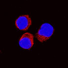
IL‑10 in Equine PBMCs.
IL-10 was detected in immersion fixed equine peripheral blood mononuclear cells (PBMCs) treated with calcium ionomycin and PMA using Goat Anti-Equine IL-10 Antigen Affinity-purified Polyclonal Antibody (Catalog # AF1605) at 15 µg/mL for 3 hours at room temperature. Cells were stained using the NorthernLights™ 557-conjugated Anti-Goat IgG Secondary Antibody (red; Catalog # NL001) and counterstained with DAPI (blue). Specific staining was localized to cytoplasm. View our protocol for Fluorescent ICC Staining of Non-adherent Cells.
Equine IL-10 ELISA Standard Curve
Recombinant Equine IL-10 (Catalog # 1605-IL) was serially diluted and captured by Goat Anti-Equine IL-10 Antigen Affinity-purified Polyclonal Antibody (Catalog # AF1605) coated on a Clear Polystyrene Microplate (Catalog # DY990). Goat Anti-Equine IL-10 Antigen Affinity-purified Polyclonal Antibody (Catalog # AF1605) was biotinylated and incubated with the protein captured on the plate. Detection of the standard curve was achieved by incubating Streptavidin-HRP (Catalog # DY998)Applications for Equine IL-10 Antibody
Application
Recommended Usage
Immunocytochemistry
10-25 µg/mL
Sample: Immersion fixed equine peripheral blood mononuclear cells
Sample: Immersion fixed equine peripheral blood mononuclear cells
Western Blot
0.1 µg/mL
Sample: Recombinant Equine IL-10 (Catalog # 1605-IL)
Sample: Recombinant Equine IL-10 (Catalog # 1605-IL)
Neutralization
Measured by its ability to neutralize IL-10-induced proliferation in the MC/9-2 mouse mast cell line. The Neutralization Dose (ND50) is typically 0.2-0.6 µg/mL in the presence of 20 ng/mL Recombinant Equine IL-10.
Equine IL-10 Sandwich Immunoassay
Please Note: Optimal dilutions of this antibody should be experimentally determined.
Formulation, Preparation, and Storage
Purification
Antigen Affinity-purified
Reconstitution
Reconstitute at 0.2 mg/mL in sterile PBS. For liquid material, refer to CoA for concentration.
Formulation
Lyophilized from a 0.2 μm filtered solution in PBS with Trehalose. *Small pack size (SP) is supplied either lyophilized or as a 0.2 µm filtered solution in PBS.
Shipping
Lyophilized product is shipped at ambient temperature. Liquid small pack size (-SP) is shipped with polar packs. Upon receipt, store immediately at the temperature recommended below.
Stability & Storage
Use a manual defrost freezer and avoid repeated freeze-thaw cycles.
- 12 months from date of receipt, -20 to -70 °C as supplied.
- 1 month, 2 to 8 °C under sterile conditions after reconstitution.
- 6 months, -20 to -70 °C under sterile conditions after reconstitution.
Background: IL-10
Interleukin 10 (IL-10), initially designated cytokine synthesis inhibitory factor (CSIF), was originally identified as a product of mouse T helper 2 (Th2) cells that inhibited the cytokine production by Th1 cells. It is a pleiotropic cytokine that regulates the immune and inflammatory responses of hematopoietic cells (1, 2). IL-10 has immunosuppressive activities and has been shown to inhibit the effector functions of monocyte/macrophage and CD4+ T cells. Conversely, IL-10 has immunostimulatory activities and can induce the proliferation and cytotoxic activity of CD8+ T cells and NK cells. IL-10 also regulates the growth and differentiation of B cells, mast cells, dendritic cells and neutrophils (1). The biological activities of IL-10 is mediated by the heteromeric IL-10 receptor complex, which is composed of the ligand-binding IL-10R alpha and the accessory IL-10R beta subunits. Both subunits belong to the class II cytokine receptor family. IL-10R beta is also utilized as a subunit in the heterodimer receptor complex for IL-22, IL-28 and IL-29. Besides IL-10, five novel cytokines (IL-19, -20, -22, -24, and -26) that share structural and limited sequence homology with IL-10 have been identified. These proteins constitute the IL-10 cytokine family (3).
Equine IL-10 cDNA encodes a 178 amino acid residue (aa) precursor protein with an 18 aa signal peptide and 160 aa mature protein that contains two potential N-linked glycosylation sites. Analogous to human IL-10, equine IL-10 likely exists as nondisulfide-linked homodimers. Equine IL-10 shares 71% and 78% aa sequence homology with mouse and human IL-10, respectively.
References
- Moore, K. et al. (2001) Annu. Rev. Immunol. 19:683.
- Mocellin, S. et al. (2003) Trends in Immunol. 23:36.
- Conti, P. et al. (2003) Immunol. Letters 88:171.
Long Name
Interleukin 10
Alternate Names
CSIF, GVHDS, IL10, IL10A, TGIF
Entrez Gene IDs
Gene Symbol
IL10
UniProt
Additional IL-10 Products
Product Documents for Equine IL-10 Antibody
Product Specific Notices for Equine IL-10 Antibody
For research use only
Loading...
Loading...
Loading...
Loading...
Loading...