Breadcrumb
- Home
- Products
- EMT Kits and Reagents
- EMT Kits and Reagents Stem Cell Products
- Human EMT 3-Color Immunocytochemistry Kit (SC026)
Human EMT 3-Color Immunocytochemistry Kit
R&D Systems, part of Bio-Techne | Catalog # SC026

Key Product Details
Summary for Human EMT 3-Color Immunocytochemistry Kit
Kit Summary
To assess epithelial to mesenchymal transition (EMT) status in human cells using single-step ICC.
Key Benefits
- Provides a straightforward readout of EMT status
- Utilizes single-step, fluorescent ICC staining of EMT markers
- Simultaneous analysis of 3 EMT markers increases confidence in EMT status
Why Assess EMT Status in Human Cells Using Established Markers?
Epithelial to mesenchymal transition (EMT) is centrally important during embryonic development and is implicated in tissue fibrosis and cancer cell metastasis. The ability to quickly and reliably assess EMT status is critical for developing a greater understanding of the mechanisms that regulate EMT.
The downregulation of epithelial markers, including E-Cadherin, and upregulation of mesenchymal markers, including Snail and Vimentin, can be used to assess EMT status.
Assessing EMT status using 3-color ICC:
- Increases confidence in EMT status by combining 3 different fluorochrome-conjugated antibodies for simultaneous staining.
- Allows researchers to evaluate changes in relative protein levels and subcellular localization of EMT markers.
- Ensures efficient use of time and reagents via single-step staining.
This kit contains the following fluorochrome-conjugated antibodies to analyze EMT status in human cells:
- NL557-conjugated Goat Anti-Human Snail
- NL637-conjugated Goat Anti-Human E-Cadherin
- NL493-conjugated Goat Anti-Human Vimentin
Each antibody is supplied as a 10X stock; enough for 25 assays when used in a 50 µL staining volume per assay.
Stability and Storage
Reagents are stable for 12 months from date of receipt when stored in the dark at 2° C to 8°.
Epithelial to Mesenchymal Transition (EMT) is a biological process which is centrally important to embryogenesis and organ development. Epithelial are highly ordered monolayers of cells with a uniform morphology. Cells within epithelial are characterized by the fact that they are adhered tightly to each other. In contrast, mesenchymal cells differ in shape and display an increased capacity for migration and invasion. This change in phenotype is thought to be involved in some oncogenic pathways. Epithelial to mesenchymal transition allows benign tumors to progress into metastatic cancers that can invade other tissues. EMT is also involved in fibrosis during scar tissue development and may be pathologically relevant to the development of progressive fibrotic diseases. Molecular markers of epithelial to mesenchymal transition include increased expression of N-Cadherin and Vimentin, nuclear localization of beta-catenin, and augmented levels of transcription factors that reduce E-Cadherin expression (i.e. Snail1).
| Species | Human |
| Source | N/A |

|
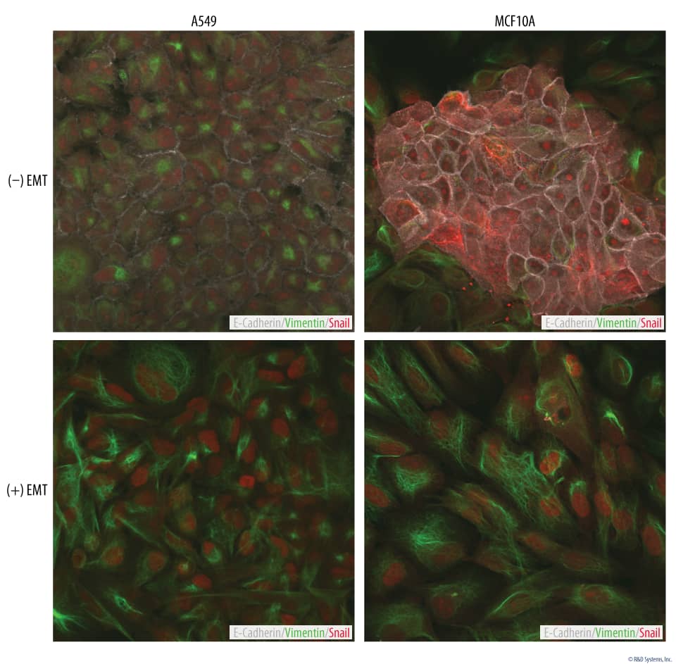
Upregulation of the Mesenchymal Markers, Snail and Vimentin, in EMT-induced Cells A549 human lung carcinoma and MCF 10A human breast epithelial cells were either untreated or cultured with the StemXVivo®EMT Inducing Media Supplement (Catalog # CCM017) for five days. The cells were analyzed for an epithelial to mesenchymal transition (EMT) by simultaneously staining with the antibodies contained in the Human EMT 3-Color Immunocytochemistry Kit (Catalog # SC026): NorthernLights™ (NL)637-conjugated Goat Anti-Human E-Cadherin (pseudocolored white), NL557-conjugated Goat Anti-Human Snail (red), and NL493-conjugated Rat Anti-Human Vimentin (green). Cells cultured with the EMT Inducing Media Supplement showed downregulation of the epithelial marker, E-Cadherin, and concurrent upregulation of the mesenchymal markers, Snail and Vimentin, compared to control cells. |
Preparation & Storage
| Shipping Conditions | The product is shipped with polar packs. Upon receipt, store it immediately at the temperature recommended below. |
| Storage | Store the unopened product at 2 - 8 °C. Do not use past expiration date. |
Assay Procedure
Refer to the product datasheet for complete product details.
Briefly, epithelial to mesenchymal transition (EMT) status in human cells can be analyzed via 3-Color ICC using the following protocol:
- EMT is induced in cells of interest
- Fluorochrome-conjugated primary antibodies are added to fixed cells
- EMT markers are visualized using a fluorescence microscope
Reagents Provided
Reagents supplied in the Human EMT 3-Color Immunocytochemistry Kit (Catalog # SC026):
- NL557-conjugated Goat Anti-Human Snail
- NL637-conjugated Goat Anti-Human E-Cadherin
- NL493-conjugated Goat Anti-Human Vimentin
This kit contains sufficient reagents to process up to 1 x 10⁹ total cells.
Other Supplies Required
Reagents
- Cells
- Coverslips (sterilized)
- Cell culture plate (24-well)
Materials
- Human pluripotent stem cells
- Pipettes and pipette tips
- Serological pipettes
Equipment
- 37 °C, 5% CO2 incubator
- Centrifuge
- Hemocytometer
- Inverted microscope
- Fluorescence microscope
Procedure Overview
Plate human epithelial cells.
Culture under desired EMT-inducing conditions.

Fix cells with 4% paraformaldehyde.

Block in blocking solution.

Incubate with fluorochrome-conjugated primary antibodies.
Wash with wash buffer.

Incubate with nuclear counterstain.

Visualize using a fluorescence microscope and appropriate filter sets

Customer Reviews for Human EMT 3-Color Immunocytochemistry Kit
There are currently no reviews for this product. Be the first to review Human EMT 3-Color Immunocytochemistry Kit and earn rewards!
Have you used Human EMT 3-Color Immunocytochemistry Kit?
Submit a review and receive an Amazon gift card!
$25/€18/£15/$25CAN/¥2500 Yen for a review with an image
$10/€7/£6/$10CAN/¥1110 Yen for a review without an image
Submit a review
