Rat Pure-Blot anti-Mouse IgG (H+L) Secondary Antibody (eB144) [DyLight 680]
Novus Biologicals, part of Bio-Techne | Catalog # NBP3-11664
![Rat Pure-Blot anti-Mouse IgG (H+L) Secondary Antibody (eB144) [DyLight 680]](https://resources.bio-techne.com/images/products/nbp3-11664_rat-pure-blot-anti-mouse-igg-h-l-secondary-antibody-eb144-dylight-680-24520231056467.jpg "Rat Pure-Blot anti-Mouse IgG (H+L) Secondary Antibody (eB144) [DyLight 680]")
![Rat Pure-Blot anti-Mouse IgG (H+L) Secondary Antibody (eB144) [DyLight 680]](https://resources.bio-techne.com/images/products/nbp3-11664_rat-pure-blot-anti-mouse-igg-h-l-secondary-antibody-eb144-dylight-680-2552023125695.jpg "Rat Pure-Blot anti-Mouse IgG (H+L) Secondary Antibody (eB144) [DyLight 680]")
Conjugate
Catalog #
Key Product Details
Species Reactivity
Mouse
Applications
Immunocytochemistry/ Immunofluorescence, Immunoprecipitation, Western Blot
Label
DyLight 680 (Excitation = 692 nm, Emission = 712 nm)
Antibody Source
Monoclonal Rat IgG Clone # eB144
Concentration
LYOPH mg/ml
Product Specifications
Immunogen
Mouse IgG
Clonality
Monoclonal
Host
Rat
Isotype
IgG
Description
This secondary antibody Conjugate was prepared from tissue culture supernatant by Protein G affinity chromatography. Assay by Immunoelectrophoresis resulted in a single precipitin arc against Anti-Mouse Serum.
Store vial at 4C prior to restoration. For extended storage aliquot contents and freeze at -20C or below. Avoid cycles of freezing and thawing. Centrifuge product if not completely clear after standing at room temperature. This product is stable for several weeks at 4C as an undiluted liquid. Dilute only prior to immediate use.
Store vial at 4C prior to restoration. For extended storage aliquot contents and freeze at -20C or below. Avoid cycles of freezing and thawing. Centrifuge product if not completely clear after standing at room temperature. This product is stable for several weeks at 4C as an undiluted liquid. Dilute only prior to immediate use.
Scientific Data Images
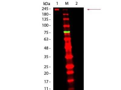
Western Blot: Rat Pure-Blot anti-Mouse IgG (H+L) Secondary Antibody (eB144) [DyLight 680] [NBP3-11664] - Western Blot of Rat Pure-Blot anti-Mouse IgG (H+L) Secondary Antibody (eB144) [DyLight 680]. Lane 1: Mouse IgG, Non-reduced. Lane 2: Mouse IgG, Reduced. Load: 50 ng per lane. Primary antibody: none. Secondary antibody: Mouse Pure-Blot anti-Rabbit IgG Secondary antibody (eB144) [DyLight 680] at 1:1,000 for 60 min at RT. Block for 30 min at RT. Predicted/Observed size: 160 kDa for Mouse IgG, Non-reduced. Migrates at slightly higher molecular weight. Other band(s): none.
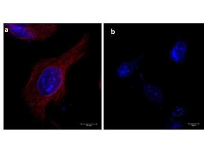
Immunocytochemistry/Immunofluorescence: Rat Pure-Blot anti-Mouse IgG (H+L) Secondary Antibody (eB144) [DyLight 680] [NBP3-11664] - Immunofluorescence microscopy of alpha-tubulin in HeLa cells using Rat Pure-Blot anti-Mouse IgG (H+L) Secondary Antibody (eB144) [DyLight 680] for detection. HeLa cells were fixed with 100% methanol, blocked (5% rat serum/0.3% Triton X-100 in 1X PBS) for 1hr, then incubated with 15ug/mL of anti-alpha-tubulin primary antibody at 4C overnight. Following 3 washes in 1X PBS for 5 min each, 5ug/mL of Rat Pure-Blot anti-Mouse IgG (H+L) Secondary Antibody (eB144) [DyLight 680] was added and allowed to incubate for 1hr at room temperature. Nuclei were counterstained with DAPI present in mounting medium. The predicted main localization is microtubules. Image taken at 63X magnification. (a) Merged alpha-tubulin (red)/DAPI (blue) image shown. (b) secondary antibody only.
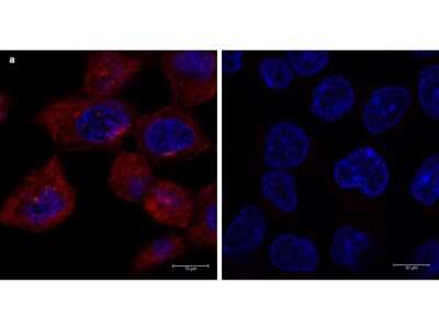
Immunocytochemistry/Immunofluorescence: Rat Pure-Blot anti-Mouse IgG (H+L) Secondary Antibody (eB144) [DyLight 680] [NBP3-11664] - Immunofluorescence microscopy of alpha-tubulin in A431 cells using Rat Pure-Blot anti-Mouse IgG (H+L) Secondary Antibody (eB144) [DyLight 680] for detection. A431 cells were fixed with 100% methanol, blocked (5% rat serum/0.3% Triton X-100 in 1X PBS) for 1 hr, then incubated with 15 ug/mL of anti-alpha-tubulin primary antibody at 4 degrees C overnight. After 3 washes in 1X PBS for 5 min each, 5 ug/mL of Rat Pure-Blot anti-Mouse IgG (H+L) Secondary Antibody (eB144) [DyLight 680] was added and allowed to incubate for 1 hr at room temperature. Nuclei were counterstained with DAPI present in mounting medium. The predicted main localization is microtubules. Image taken at 63X magnification. (a) Merged alpha-tubulin (red)/DAPI (blue) image shown. (b) secondary antibody only.
Applications
Application
Recommended Usage
Immunocytochemistry/ Immunofluorescence
1:500 - 1:2500
Western Blot
1:1000
Application Notes
This antibody has been tested in ELISA, immunofluorescence, immunoprecipitation, and western blot and may also be used for detection in immunoassays that do not employ immunoprecipitation. This antibody is provided as a lyophilized powder. To conserve reagent, we recommend incubating the blots from minigels in sealed bags (removing as much air as possible) with minimal volume (2-3 mLs). If used conservatively at 2.5mls/blot will yield enough reagent for 40 blots. Note that there are three key procedural considerations: 1. Protein A or G beads may be used with the mouse, goat and sheep Pure-Blot secondaries, but not with the rabbit Pure-Blot secondary. Use of protein A or G beads with the rabbit Pure-Blot will result in contaminating bands. 2. Immunoprecipitate should be completely reduced. 3. blocking buffer for Fluorescent Western Blotting should be used as the blocking protein for the immunoblot. Note: To achieve best results when detecting mouse IgG1 subtypes, we recommend performing a dot blot or titration to determine the optimal dilution factor for your desired application. All recommended dilutions for listed applications are intended as an initial recommendation, specific conditions for each protein and antibody combination should be specifically optimized by the end user.
Formulation, Preparation, and Storage
Purification
Protein G purified
Reconstitution
Reconstitute with 100 ul deionized water (or equivalent).
Formulation
Lyophilized from 0.02 M Potassium Phosphate, 0.15 M Sodium Chloride, pH 7.2, 10 mg/ml Polyethylene Glycol (PEG-8000)
Preservative
0.01% Sodium Azide
Concentration
LYOPH mg/ml
Shipping
The product is shipped with polar packs. Upon receipt, store it immediately at the temperature recommended below.
Stability & Storage
Store lyophilized antibody at 4C. Aliquot reconstituted liquid and store at -20C. Avoid freeze-thaw cycles.
Background: IgG (H+L)
The 4 IgG subclasses, sharing 95% amino acid identity, include IgG1, IgG2, IgG3, and IgG4 for humans and IgG1, IgG2a, IgG2b, and IgG3 for mice. The relative abundance of each human subclass is 60% for IgG1, 32% for IgG2, 4% for IgG3, and 4% for IgG4. In an IgG deficiency, there may be a shortage of one or more subclasses (4).
References
1. Painter RH. (1998) Encyclopedia of Immunology (Second Edition). Elsevier. 1208-1211
2. Chapter 9 - Antibodies. (2012) Immunology for Pharmacy. Mosby 70-78
3. Schroeder H, Cavacini, L. (2010) Structure and Function of Immunoglobulins. J Allergy Clin Immunol. 125(2 0 2): S41-S52. PMID: 20176268
4. Vidarsson G, Dekkers G, Rispens T. (2014) IgG subclasses and allotypes: from structure to effector functions. Front Immunol. 5:520. PMID: 25368619
Alternate Names
IP Detection Reagent
Additional IgG (H+L) Products
Product Specific Notices
DyLight (R) is a trademark of Thermo Fisher Scientific Inc. and its subsidiaries.
This product is for research use only and is not approved for use in humans or in clinical diagnosis. Secondary Antibodies are guaranteed for 1 year from date of receipt.
Loading...
Loading...
Loading...
Loading...
Loading...