
Mouse VEGF Quantikine ELISA Kit
R&D Systems, part of Bio-Techne | Catalog # MMV00

Key Product Details
Assay Length
4.5 hours
Sample Type & Volume Required Per Well
Cell Culture Supernates (50 µL), Tissue Homogenates (50 µL), Serum (10 µL), EDTA Plasma (10 µL), Heparin Plasma (10 µL)
Sensitivity
3 pg/mL
Assay Range
7.8-500 pg/mL (Cell Culture Supernates, Tissue Homogenates, Serum, EDTA Plasma)
Product Summary for Mouse VEGF Quantikine ELISA Kit
The Quantikine Mouse VEGF Immunoassay is a 4.5 hour solid phase ELISA designed to measure mouse VEGF in cell culture supernates, tissue homogenates, mouse serum, and plasma. It contains Sf 21-expressed mouse VEGF and antibodies raised against the recombinant factor. This immunoassay has been shown to quantitate the recombinant mouse VEGF accurately. Results obtained using natural mouse VEGF showed dose-response curves that were parallel to the standard curves obtained using the recombinant Quantikine kit standards. These results indicate that this kit can be used to determine relative mass values for natural mouse VEGF.
Product Specifications
Assay Type
Solid Phase Sandwich ELISA
Format
96-well strip plate
Measurement
Quantitative ELISA
Detection Method
Colorimetric - 450nm (TMB)
Conjugate
HRP
Reactivity
Mouse
Specificity
This kit recognizes both the 164 and 120 amino acid residue forms of mouse VEGF
Cross-reactivity
< 0.5% cross-reactivity observed with available related molecules. < 50% cross-species reactivity observed with species tested.
Interference
Interference observed with 1 or more available related molecules.
Precision
Intra-Assay Precision (Precision within an assay) Three samples of known concentration were tested on one plate to assess intra-assay precision.
Inter-Assay Precision (Precision between assays) Three samples of known concentration were tested in separate assays to assess inter-assay precision.
Cell Culture Supernates, EDTA Plasma, Serum, Tissue Homogenates
| Intra-Assay Precision | Inter-Assay Precision | |||||
|---|---|---|---|---|---|---|
| Sample | 1 | 2 | 3 | 1 | 2 | 3 |
| n | 20 | 20 | 20 | 20 | 20 | 20 |
| Mean (pg/mL) | 37.7 | 144 | 277 | 38.3 | 138 | 291 |
| Standard Deviation | 3.1 | 6.2 | 12.9 | 3.2 | 7.8 | 18.5 |
| CV% | 8.2 | 4.3 | 4.7 | 8.4 | 5.7 | 6.4 |
Recovery for Mouse VEGF Quantikine ELISA Kit
The recovery of mouse VEGF spiked to three levels throughout the range of the assay in various matrices was evaluated.
| Sample Type | Average % Recovery | Range % |
|---|---|---|
| Cell Culture Supernates (n=9) | 103 | 95-114 |
| EDTA Plasma (n=5) | 91 | 82-103 |
| Heparin Plasma (n=4) | 89 | 87-94 |
| Serum (n=9) | 101 | 83-115 |
| Tissue Homogenates (n=3) | 91 | 76-107 |
Linearity
To assess the linearity of the assay, samples containing and/or spiked with various concentrations of mouse VEGF in each matrix were diluted with Calibrator Diluent and then assayed.

Scientific Data Images for Mouse VEGF Quantikine ELISA Kit

Mouse VEGF ELISA Standard Curve
Mouse VEGF Quantikine ELISA Kit
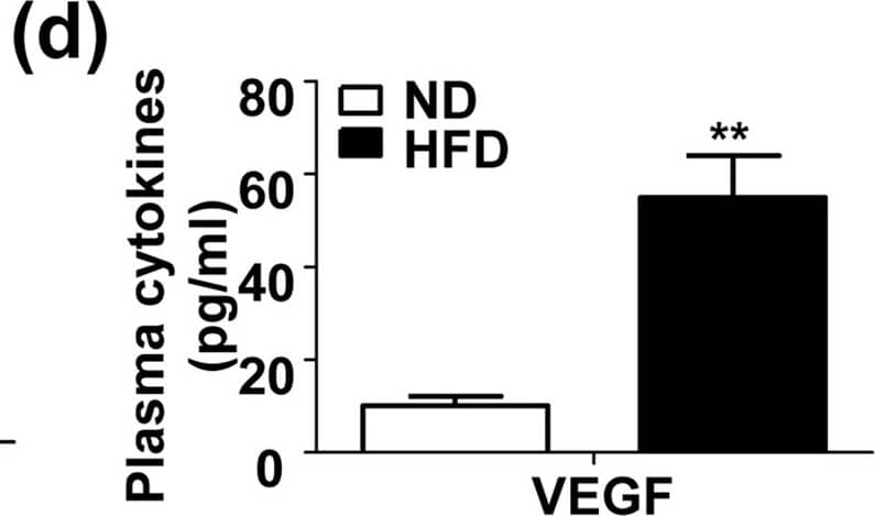
Increased production of proinflammatory cytokines and VEGF in the plasma and chondrocytes in high fat diet (HFD)-feeding mice.(a~d) levels of plasma IL-6 (a), TNF-alpha (b), IL-1 beta (c) and VEGF (d) in the C57BL/6 male mice fed with a normal diet (ND) or HFD for 8 weeks. (n = 5, **P < 0.01). (e~h) Relative mRNA expression of IL-6 (e), TNF-alpha (f), IL-1 beta (g) and VEGF (h) in the chondrocytes isolated from the 8-week ND or HFD-feeding mice. (n = 5, **P < 0.01). Image collected and cropped by CiteAb from the following open publication (https://pubmed.ncbi.nlm.nih.gov/26271607), licensed under a CC-BY license. Not internally tested by R&D Systems.
Detection of Mouse VEGF
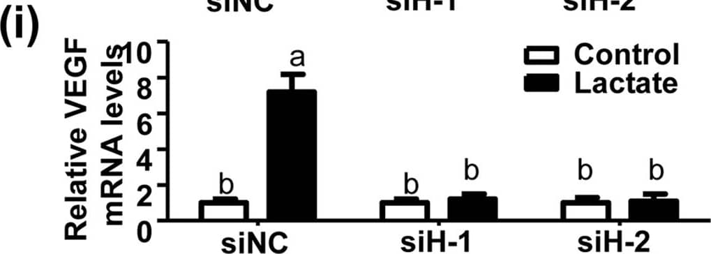
HFD-induced plasma saturated free fatty acid (FFA) and lactate trigger production of proinflammatory cytokines and VEGF in mouse chondrocytes.(a~d) Relative mRNA levels of IL-6 (a), TNF-alpha (b), IL-1 beta (c) and VEGF (d) in the mouse primary chondrocytes treated with full or fractionated (>3 KD or <3 KD) serum from the 8-week ND or HFD-feeding mice. (n = 3, *P < 0.05, **P < 0.01 and ***P < 0.005). (e~h) Relative mRNA levels of IL-6 (e), TNF-alpha (f), IL-1 beta (g) and VEGF (h) in the mouse primary chondrocytes treated with BSA (5%) or BSA-linked FFA (18:0, 200 μM) for 24 h. (n = 3, *P < 0.05 and **P < 0.01). (i~l) Relative mRNA expression of IL-6 (i), TNF-alpha (j), IL-1 beta (k) and VEGF (l) in the mouse primary chondrocytes treated with lactate (25 mM) or vehicle control for 24 h. (n = 3, *P < 0.05 and **P < 0.01). Image collected and cropped by CiteAb from the following open publication (https://pubmed.ncbi.nlm.nih.gov/26271607), licensed under a CC-BY license. Not internally tested by R&D Systems.Preparation and Storage
Shipping
The product is shipped at ambient temperature. Upon receipt, store it immediately at the temperature recommended below.
Stability & Storage
Store the unopened product at 2 - 8 °C. Do not use past expiration date.
Background: VEGF
VEGF dimers bind to two related receptor tyrosine kinases, VEGF R1 (also called Flt-1) and
VEGF R2 (Flk-1/KDR), and induce their homodimerization and autophosphorylation (3, 4, 7, 17,
18). These receptors have seven extracellular immunoglobulin-like domains and an intracellular
split tyrosine kinase domain. They are expressed on vascular endothelial cells and a range of
non-endothelial cells. Although VEGF affinity is highest for binding to VEGF R1, VEGF R2
appears to be the primary mediator of VEGF angiogenic activity (3, 4). VEGF165 also binds the
semaphorin receptor, neuropilin-1, which promotes complex formation with VEGF R2 (19).
VEGF is best known for its role in vasculogenesis. During embryogenesis, VEGF regulates the
proliferation, migration, and survival of endothelial cells (3, 4), thus regulating blood vessel
density and size but playing no role in determining vascular patterns. VEGF promotes bone
formation through osteoblast and chondroblast recruitment and is also a monocyte
chemoattractant (20-22). In postnatal life, VEGF maintains endothelial cell integrity and is a
potent mitogen for micro- and macro-vascular endothelial cells. In adults, VEGF functions
mainly in wound healing and the female reproductive cycle (3). In diseased tissues, VEGF
promotes vascular permeability. It is thus thought to contribute to tumor metastasis by
promoting both extravasation and tumor angiogenesis (23, 24). Various strategies have been
employed therapeutically to antagonize VEGF-mediated tumor angiogenesis (25). Circulating
VEGF levels correlate with disease activity in autoimmune diseases such as rheumatoid
arthritis, multiple sclerosis, and systemic lupus erythematosus (26).
Long Name
Vascular Endothelial Growth Factor
Alternate Names
MVCD1, VAS, Vasculotropin, VEGF-A, VEGFA, VPF
Entrez Gene IDs
Gene Symbol
VEGFA
Additional VEGF Products
Product Documents for Mouse VEGF Quantikine ELISA Kit
Product Specific Notices for Mouse VEGF Quantikine ELISA Kit
For research use only
⚠ WARNING: This product can expose you to chemicals including N,N-Dimethylforamide, which is known to the State of California to cause cancer. For more information, go to www.P65Warnings.ca.gov.
Loading...
Loading...
Loading...
Loading...