
OPA1 Antibody (1E8-1D9) - BSA Free
Novus Biologicals, part of Bio-Techne | Catalog # NBP1-71656

![Simple Western: OPA1 Antibody (1E8-1D9)BSA Free [NBP1-71656]](https://resources.bio-techne.com/images/products/OPA1-Antibody-1E8-1D9-Simple-Western-NBP1-71656-img0010.jpg "Simple Western: OPA1 Antibody (1E8-1D9)BSA Free [NBP1-71656]")
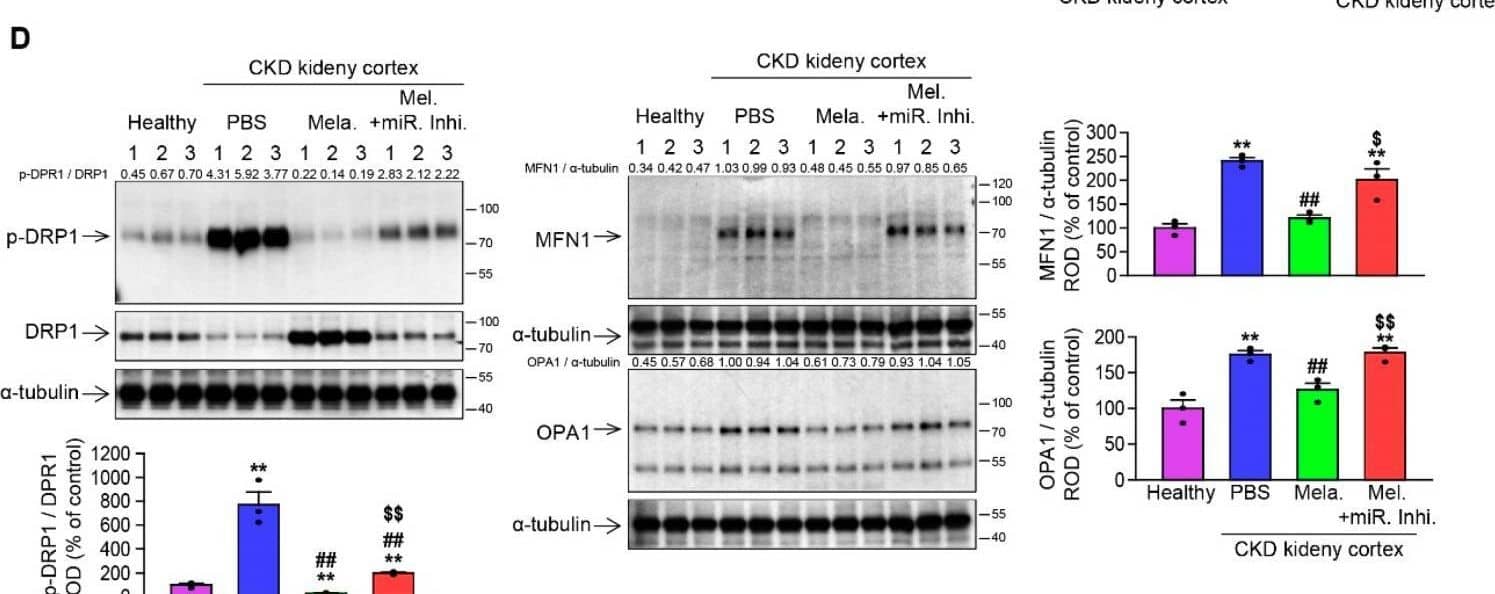
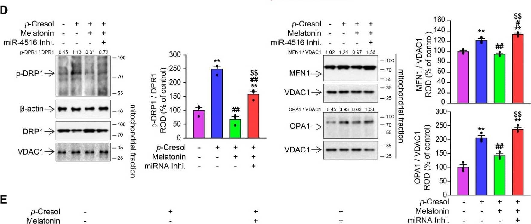
![Western Blot: OPA1 Antibody (1E8-1D9)BSA Free [NBP1-71656]](https://resources.bio-techne.com/images/products/OPA1-Antibody-1E8-1D9-Western-Blot-NBP1-71656-img0009.jpg "Western Blot: OPA1 Antibody (1E8-1D9)BSA Free [NBP1-71656]")
Scientific images may be resized, cropped, or adjusted for brightness or contrast for presentation purposes. These updates are limited to presentation and do not affect the underlying scientific interpretation of the data. Where available, additional source data may be provided upon request.
Conjugate
Catalog #
Key Product Details
Species Reactivity
Validated:
Human, Mouse, Rat, Bovine, Chinese Hamster
Cited:
Human, Mouse
Applications
Validated:
Immunohistochemistry, Immunohistochemistry-Paraffin, Western Blot, Immunocytochemistry/ Immunofluorescence, Simple Western
Cited:
Western Blot, Simple Western, IF/IHC
Label
Unconjugated
Antibody Source
Monoclonal Mouse IgG1 kappa Clone # 1E8-1D9
Format
BSA Free
Concentration
1.0 mg/ml
Product Specifications
Immunogen
Human OPA1 [Swiss-Prot# O60313].
Localization
Mitochondrion inner membrane; Single-pass membrane protein. Mitochondrion intermembrane space
Clonality
Monoclonal
Host
Mouse
Isotype
IgG1 kappa
Scientific Data Images for OPA1 Antibody (1E8-1D9) - BSA Free
Western Blot: OPA1 Antibody (1E8-1D9)BSA Free [NBP1-71656]
Western Blot: OPA1 Antibody (1E8-1D9) [NBP1-71656] - Analysis of OPA1 expression in 1) HeLa 2) MEF 3) HepG2 4) A431 5) CHO 6)PC12 and 7) Ntera2 whole cell lysates using NBP1-71656.![Immunocytochemistry/ Immunofluorescence: OPA1 Antibody (1E8-1D9) - BSA Free [NBP1-71656]](https://resources.bio-techne.com/images/products/OPA1-Antibody-1E8-1D9-Immunocytochemistry-Immunofluorescence-NBP1-71656-img0007.jpg "Immunocytochemistry/ Immunofluorescence: OPA1 Antibody (1E8-1D9) - BSA Free [NBP1-71656]")
Immunocytochemistry/ Immunofluorescence: OPA1 Antibody (1E8-1D9) - BSA Free [NBP1-71656]
Immunocytochemistry/Immunofluorescence: OPA1 Antibody (1E8-1D9) [NBP1-71656] - OPA1 antibody was tested in ARPE-19 cells with FITC (green). Nuclei and actin were counterstained with DAPI (blue) and Phalloidin (red).![Immunohistochemistry: OPA1 Antibody (1E8-1D9) - BSA Free [NBP1-71656]](https://resources.bio-techne.com/images/products/OPA1-Antibody-1E8-1D9-Immunohistochemistry-NBP1-71656-img0008.jpg "Immunohistochemistry: OPA1 Antibody (1E8-1D9) - BSA Free [NBP1-71656]")
Immunohistochemistry: OPA1 Antibody (1E8-1D9) - BSA Free [NBP1-71656]
Immunohistochemistry: OPA1 Antibody (1E8-1D9) [NBP1-71656] - Analysis of OPA1 on mouse skin using NBP1-71656.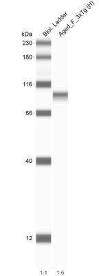
Applications for OPA1 Antibody (1E8-1D9) - BSA Free
Application
Recommended Usage
Immunocytochemistry/ Immunofluorescence
1:50
Immunohistochemistry
1:100
Immunohistochemistry-Paraffin
1:100
Simple Western
1:25
Western Blot
1:1000
Application Notes
In Western blot, multiple protein isoforms can be seen at ~90, 80 and 65 kDa.
In Simple Western only 10 - 15 uL of the recommended dilution is used per data point.
See Simple Western Antibody Database for Simple Western validation: Tested in HeLa lysate 1.0 mg/mL, separated by Size, antibody dilution of 1:25, apparent MW was 93 kDa. Separated by Size-Wes, Sally Sue/Peggy Sue.
In Simple Western only 10 - 15 uL of the recommended dilution is used per data point.
See Simple Western Antibody Database for Simple Western validation: Tested in HeLa lysate 1.0 mg/mL, separated by Size, antibody dilution of 1:25, apparent MW was 93 kDa. Separated by Size-Wes, Sally Sue/Peggy Sue.
Reviewed Applications
Read 2 reviews rated 5 using NBP1-71656 in the following applications:
Formulation, Preparation, and Storage
Purification
Protein G purified
Formulation
PBS
Format
BSA Free
Preservative
0.02% Sodium Azide
Concentration
1.0 mg/ml
Shipping
The product is shipped with polar packs. Upon receipt, store it immediately at the temperature recommended below.
Stability & Storage
Store at 4C short term. Aliquot and store at -20C long term. Avoid freeze-thaw cycles.
Background: OPA1
Long Name
Optic Atrophy Protein 1
Alternate Names
BERHS, EC 3.6.5.5, LargeG, lilr3, MGM1, MTDPS14, NPG, NTG
Gene Symbol
OPA1
UniProt
Additional OPA1 Products
Product Documents for OPA1 Antibody (1E8-1D9) - BSA Free
Product Specific Notices for OPA1 Antibody (1E8-1D9) - BSA Free
This product is for research use only and is not approved for use in humans or in clinical diagnosis. Primary Antibodies are guaranteed for 1 year from date of receipt.
Loading...
Loading...
Loading...
Loading...