
Mouse S100A4 Antibody
R&D Systems, part of Bio-Techne | Catalog # MAB11727
Recombinant Monoclonal Antibody.

Conjugate
Catalog #
Key Product Details
Species Reactivity
Mouse
Applications
Multiplex Immunofluorescence, Immunohistochemistry, Western Blot, Immunocytochemistry, Simple Western, COMET
Label
Unconjugated
Antibody Source
Recombinant Monoclonal Rabbit IgG Clone # 3352C
Product Specifications
Immunogen
E. coli-derived mouse S100A4
Leu20-Pro219
Accession # P07091
Leu20-Pro219
Accession # P07091
Specificity
Detects recombinant mouse S100A4 protein in Direct ELISA.
Clonality
Monoclonal
Host
Rabbit
Isotype
IgG
Scientific Data Images for Mouse S100A4 Antibody
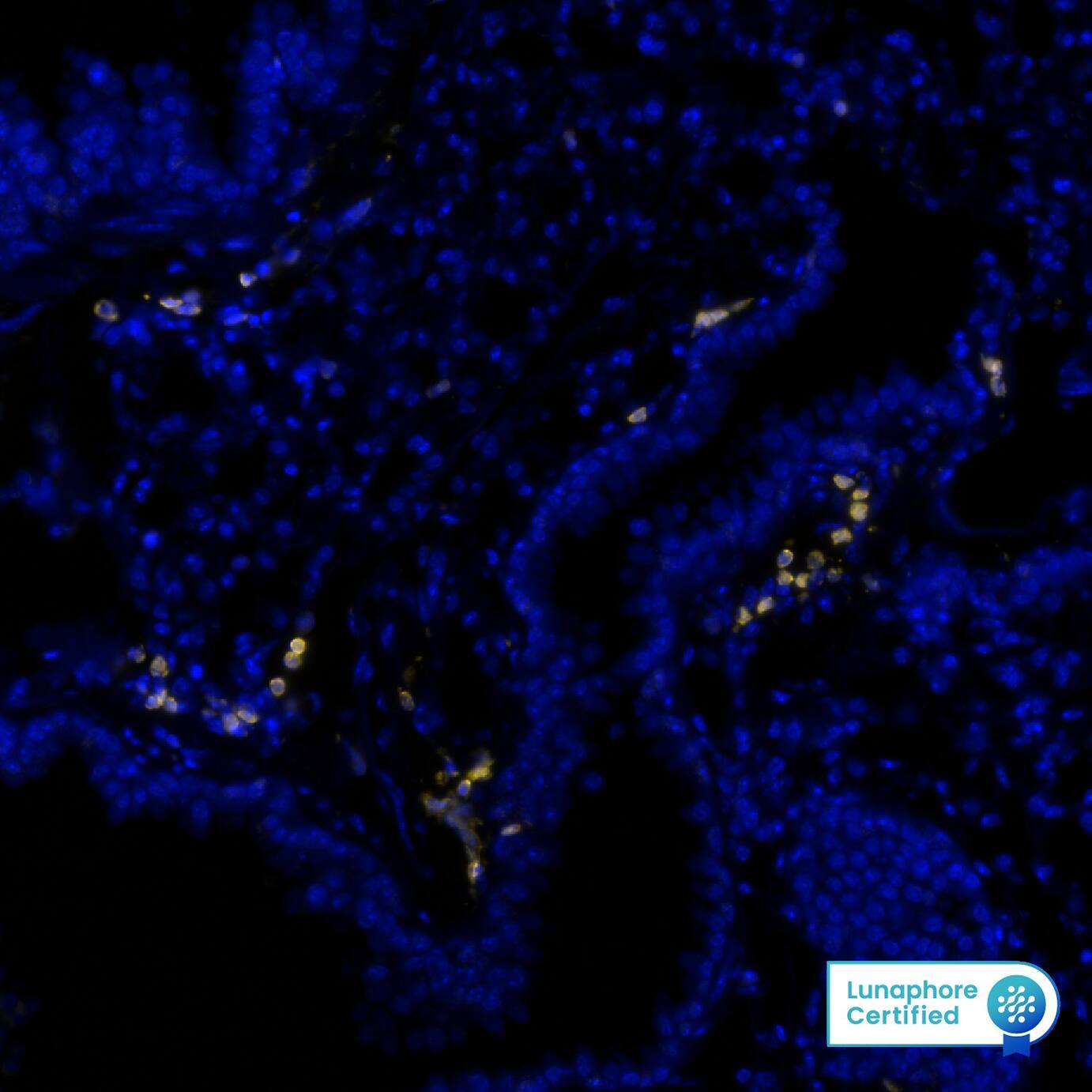
Detection of S100A4 in Mouse Lung via seqIF™ staining on COMET™
S100A4 was detected in perfusion fixed paraffin-embedded sections of mouse Lung using Rabbit Anti-Mouse S100A4, Monoclonal Antibody (Catalog # MAB11727) at 0.1ug/mL at 37° Celsius for 4 minutes. Before incubation with the primary antibody, tissue underwent an all-in-one dewaxing and antigen retrieval preprocessing using PreTreatment Module (PT Module) and Dewax and HIER Buffer H (pH 9; Epredia Catalog # TA-999-DHBH). Tissue was stained using the Alexa Fluor™ Plus 647 Goat anti-Rabbit IgG Secondary Antibody at 1:200 at 37 ° Celsius for 2 minutes. (Yellow; Lunaphore Catalog # DR647RB) and counterstained with DAPI (blue; Lunaphore Catalog # DR100). Specific staining was localized to the nucleus and cytoplasm. Protocol available in COMET™ Panel Builder.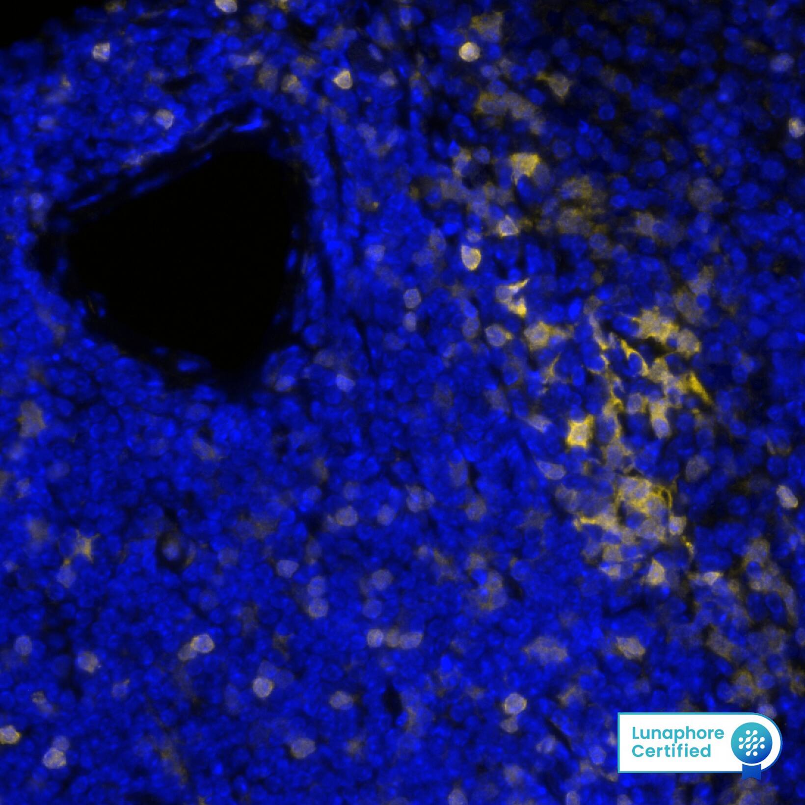
Detection of S100A4 in Mouse Spleen via seqIF™ staining on COMET™
S100A4 was detected in immersion fixed paraffin-embedded sections of mouse Spleen using Rabbit Anti-Mouse S100A4, Monoclonal Antibody (Catalog # MAB11727) at 0.1ug/mL at 37° Celsius for 4 minutes. Before incubation with the primary antibody, tissue underwent an all-in-one dewaxing and antigen retrieval preprocessing using PreTreatment Module (PT Module) and Dewax and HIER Buffer H (pH 9; Epredia Catalog # TA-999-DHBH). Tissue was stained using the Alexa Fluor™ Plus 647 Goat anti-Rabbit IgG Secondary Antibody at 1:200 at 37 ° Celsius for 2 minutes. (Yellow; Lunaphore Catalog # DR647RB) and counterstained with DAPI (blue; Lunaphore Catalog # DR100). Specific staining was localized to the nucleus and cytoplasm. Protocol available in COMET™ Panel Builder.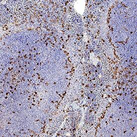
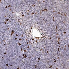
Detection of S100A4 in Mouse Spleen.
S100A4 was detected in perfusion fixed paraffin-embedded sections of mouse spleen using Rabbit Anti-Mouse S100A4 Monoclonal Antibody (Catalog # MAB11727) at 0.05 µg/ml for 1 hour at room temperature followed by incubation with the Anti-Rabbit IgG VisUCyte™ HRP Polymer Antibody (Catalog # VC003) or the HRP-conjugated Anti-Rabbit IgG Secondary Antibody (Catalog # HAF008). Before incubation with the primary antibody, tissue was subjected to heat-induced epitope retrieval using VisUCyte Antigen Retrieval Reagent-Basic (Catalog # VCTS021). Tissue was stained using DAB (brown) and counterstained with hematoxylin (blue). Specific staining was localized to the nucleus and cytoplasm. View our protocol for IHC Staining with VisUCyte HRP Polymer Detection Reagents.
Applications for Mouse S100A4 Antibody
Application
Recommended Usage
COMET
Immunocytochemistry
0.3-3 µg/mL
Sample: S100A4 was detected in Immersion fixed RAW264.7 cells
Sample: S100A4 was detected in Immersion fixed RAW264.7 cells
Immunohistochemistry
0.05-10 µg/mL
Sample: Perfusion fixed paraffin-embedded sections of mouse spleen and thymus
Sample: Perfusion fixed paraffin-embedded sections of mouse spleen and thymus
Multiplex Immunofluorescence
0.1 µg/mL
Sample: Perfusion fixed paraffin-embedded sections of mouse Spleen and Lung
Sample: Perfusion fixed paraffin-embedded sections of mouse Spleen and Lung
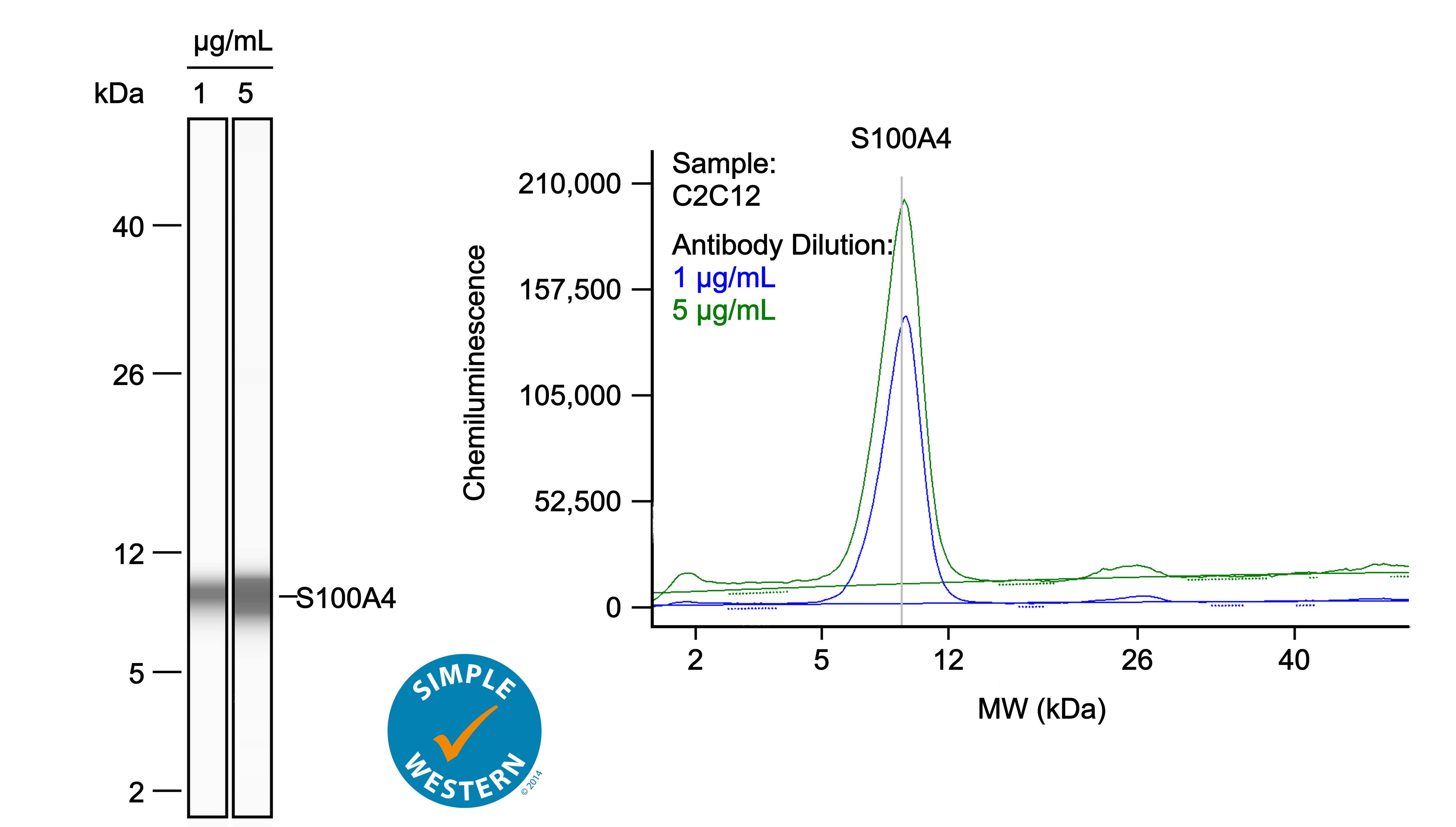
Simple Western
1-5 µg/mL
Sample: C2C12 mouse myoblast cell line
Sample: C2C12 mouse myoblast cell line
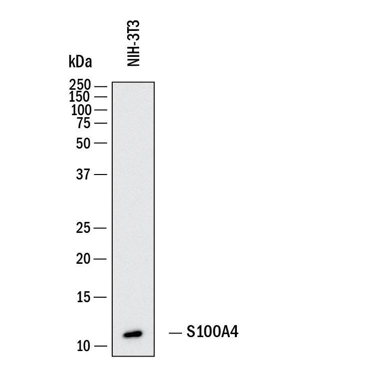
Western Blot
0.05 µg/mL
Sample: NIH-3T3 mouse embryonic fibroblast cell line
Sample: NIH-3T3 mouse embryonic fibroblast cell line
Formulation, Preparation, and Storage
Purification
Protein A or G purified from hybridoma culture supernatant
Reconstitution
Reconstitute lyophilized material at 0.2 mg/ml in sterile PBS. For liquid material, refer to CoA for concentration.
Formulation
Lyophilized from a 0.2 μm filtered solution in PBS with Trehalose.
Shipping
Lyophilized product is shipped at ambient temperature. Liquid small pack size (-SP) is shipped with polar packs. Upon receipt, store immediately at the temperature recommended below.
Stability & Storage
Use a manual defrost freezer and avoid repeated freeze-thaw cycles.
- 12 months from date of receipt, -20 to -70 °C as supplied.
- 1 month, 2 to 8 °C under sterile conditions after reconstitution.
- 6 months, -20 to -70 °C under sterile conditions after reconstitution.
Background: S100A4
Mouse S100 calcium-binding protein A4 (S100A4; also metastasin, mts1, metastatic cell protein, and placental calcium-binding protein) is a 12 kDa member of the S100 family of calcium-binding proteins. S100A4 is 101 amino acids (aa) in length and contains two calcium-binding EF-hand domains (aa 12 - 47 and 50 - 85). Mouse S100A4 shares 96%, 93%, and 90% aa sequence identity with rat, human, and bovine S100A4, respectively. S100A4 is involved in regulating the metastatic behavior of tumor cells. It is ubiquitously expressed in tumor cells and normal cells, but the level of S100A4 expression correlates with the metastatic potential of tumor cells.
Long Name
S100 Calcium Binding Protein A4
Alternate Names
18A2, 42A, CAPL, MTS1, P9KA, PEL98
Gene Symbol
S100A4
UniProt
Additional S100A4 Products
Product Documents for Mouse S100A4 Antibody
Product Specific Notices for Mouse S100A4 Antibody
For research use only
Loading...
Loading...
Loading...
Loading...
Loading...