Mouse S100A4 Antibody
R&D Systems, part of Bio-Techne | Catalog # MAB11673


Key Product Details
Species Reactivity
Applications
Label
Antibody Source
Product Specifications
Immunogen
Ala2-Lys101
Accession # P07091
Specificity
Clonality
Host
Isotype
Scientific Data Images for Mouse S100A4 Antibody
S100A4 in Mouse Spleen via seqIF™ staining on COMET™
S100A4 was detected in immersion fixed paraffin-embedded sections of mouse Spleen using Rat Anti-Mouse S100A4, Monoclonal Antibody (Catalog #MAB11673) at 0.5ug/mL at 32° Celsius for 4 minutes. Before incubation with the primary antibody, tissue underwent an all-in-one dewaxing and antigen retrieval preprocessing using PreTreatment Module (PT Module) and Dewax and HIER Buffer H (pH 9; Epredia Catalog # TA-999-DHBH). Tissue was stained using the Alexa Fluor™ 555 Goat anti-Rat IgG Secondary Antibody at 1:100 at 37 ° Celsius for 2 minutes. (Yellow; Lunaphore Catalog # DR555RT) and counterstained with DAPI (blue; Lunaphore Catalog # DR100). Specific staining was localized to the cytoplasm and nucleus. Protocol available in COMET™ Panel Builder.Detection of Mouse S100A4 by Western Blot.
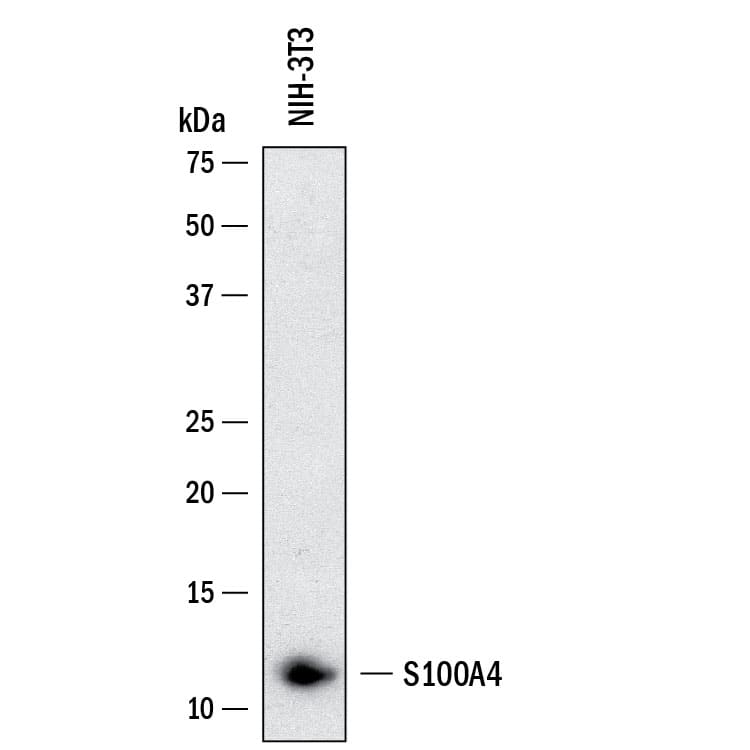
Western Blot shows lysates of NIH-3T3 mouse embryonic fibroblast cell line. PVDF membrane was probed with 1 µg/ml of Rat Anti-Mouse S100A4 Monoclonal Antibody (Catalog # MAB11673) followed by HRP-conjugated Anti-Rat IgG Secondary Antibody (Catalog # HAF005). A specific band was detected for S100A4 at approximately 11 kDa (as indicated). This experiment was conducted under reducing conditions and using Western Blot Buffer Group 1.Detection of S100A4 in Mouse Spleen.
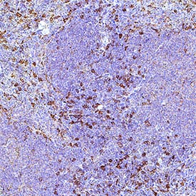
S100A4 was detected in immersion fixed paraffin-embedded sections of mouse spleen using Rat Anti-Mouse S100A4 Monoclonal Antibody (Catalog # MAB11673) at 5 µg/ml overnight at 4 °C. Before incubation with the primary antibody, tissue was subjected to heat-induced epitope retrieval using VisUCyte Antigen Retrieval Reagent-Basic (Catalog # VCTS021). Tissue was stained using the HRP-conjugated Anti-Rat IgG Secondary Antibody (Catalog # HAF005) and counterstained with hematoxylin (blue). Specific staining was localized to the nucleus and cytoplasm. View our protocol for Chromogenic IHC Staining of Paraffin-embedded Tissue Sections.Applications for Mouse S100A4 Antibody
Immunohistochemistry
Sample: Immersion fixed paraffin-embedded sections of mouse spleen
Multiplex Immunofluorescence
Sample: Immersion fixed paraffin-embedded sections of mouse spleen
Simple Western
Sample: NIH-3T3 mouse embryonic fibroblast cell line
Western Blot
Sample: NIH-3T3 mouse embryonic fibroblast cell line
Formulation, Preparation, and Storage
Purification
Reconstitution
Formulation
Shipping
Stability & Storage
- 12 months from date of receipt, -20 to -70 °C as supplied.
- 1 month, 2 to 8 °C under sterile conditions after reconstitution.
- 6 months, -20 to -70 °C under sterile conditions after reconstitution.
Background: S100A4
S100A4 (also named Metastasin, Mtsl and Calvasculin) is an 11 kDa member of the S100 (soluble in 100% saturated ammonium sulfate) family of proteins (1‑5). S100 family members belong to the EF-hand superfamily of Ca++-binding proteins. These participate in both calcium‑dependent and calcium‑independent protein‑protein interactions. The hallmark of this superfamily is the EF-hand motif that consists of a Ca++-binding site flanked by two alpha-helices (helix E and helix F) that were originally identified in a right-handed model of carp muscle calcium‑binding protein (6). Mouse S100A4 is 101 amino acids (aa) in length (1, 2). It contains two EF hand domains (aa 12‑47 and aa 50‑85). The first domain has a 14 aa cation-binding motif and binds Ca++ with low affinity. The second Ca++-binding motif is 12 aa in length and binds Ca++ with high affinity. S100A4 has no classical signal sequence but is secreted from cells (3, 7). Mouse S100A4 shares 93%, 96% and 89% aa identity with human, rat and canine S100A4, respectively. S100A4 exists as dimer (8, 9, 10). Extracellular S100A4 is reported to induce MMP production, activate MMPs, promote neurite outgrowth and stimulate cardiomyocyte proliferation (4, 10, 11, 12, 13). Within the cell, dimers are likely the functional unit. Here, they are constitutive homo‑ or heterodimers (with S100A1) that interact with Ca++, undergo a conformational change, and subsequently bind to cytoplasmic targets. Known targets include p53, Myosin heavy chain II, F-actin and Liprin beta1 (4, 14). In general, it can be said that S100A4 blocks target phosphorylation and multimerization (4, 7, 14). S100A4 activity has been associated with cell transformation. It seems likely this is either coincidental, or a consequence, rather than a cause of transformation (3).
References
- Jackson-Grusby, L.L. et al. (1987) Nucleic Acids Res. 15:6677.
- Goto, K. et al. (1988) J. Biochem. 103:48.
- Garrett, S.C. et al. (2006) J. Biol. Chem. 281:677.
- Santamaria-Kisiel, L. et al. (2006) Biochem. J. 396:201.
- Donato, R. (2001) Int. J. Biochem. Mol. Biol. 33:637.
- Kretsinger, R.H. and C.E. Nockolds (1973) J. Biol. Chem. 248:3313.
- Helfman, D.M. et al. (2005) Br. J. Cancer 92:1955.
- Burkitt, W.I. et al. (2003) Biochem. Soc. Trans. 31:985.
- Vallaly, K.M. et al. (2002) Biochemistry 41:12670.
- Novitskaya, V. et al. (2000) J. Biol. Chem. 275:41278.
- Stary, M. et al. (2006) Biochem. Biophys. Res. Commun. 343:555.
- Semov, A. et al. (2005) J. Biol. Chem. 280:20833.
- Saleem, M. et al. (2006) Proc. Natl. Acad. Sci. USA 103:14825.
- Kriajevska, M. et al. (2002) J. Biol. Chem. 277:5229.
Long Name
Alternate Names
Gene Symbol
UniProt
Additional S100A4 Products
Product Documents for Mouse S100A4 Antibody
Product Specific Notices for Mouse S100A4 Antibody
For research use only


