
Mouse NCAM-1/CD56 Antibody
R&D Systems, part of Bio-Techne | Catalog # MAB7820

Scientific images may be resized, cropped, or adjusted for brightness or contrast for presentation purposes. These updates are limited to presentation and do not affect the underlying scientific interpretation of the data. Where available, additional source data may be provided upon request.
Conjugate
Catalog #
Key Product Details
Species Reactivity
Validated:
Mouse
Cited:
Human, Mouse
Applications
Validated:
Multiplex Immunofluorescence, Immunohistochemistry, Western Blot, Flow Cytometry, COMET, CyTOF-ready
Cited:
Western Blot, Flow Cytometry
Label
Unconjugated
Antibody Source
Monoclonal Rat IgG2A Clone # 809220
Product Specifications
Immunogen
Mouse myeloma cell line NS0-derived recombinant mouse NCAM‑1/CD56
Leu20-Thr711
Accession # P13595
Leu20-Thr711
Accession # P13595
Specificity
Detects mouse NCAM‑1/CD56 in direct ELISAs and Western blots.
Clonality
Monoclonal
Host
Rat
Isotype
IgG2A
Scientific Data Images for Mouse NCAM-1/CD56 Antibody
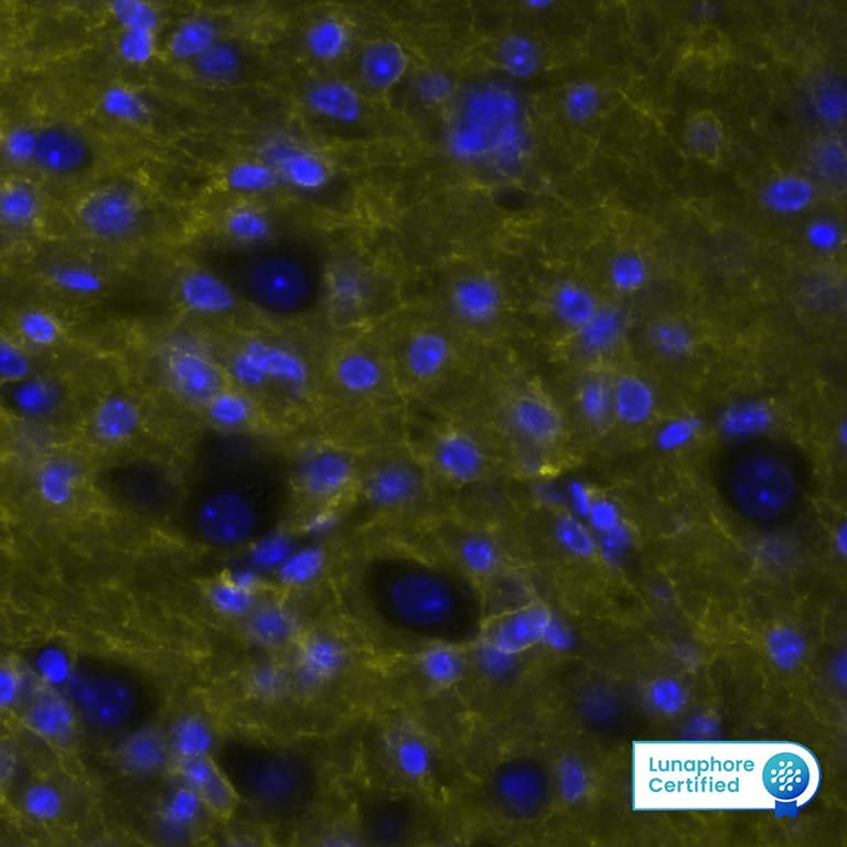
Detection of CD56/NCAM1 in Mouse Cerebellum via seqIF™ staining on COMET™
CD56/NCAM1 was detected in immersion fixed paraffin-embedded sections of mouse Cerebellum using Rat Anti-Mouse CD56, Monoclonal Antibody (Catalog # MAB7820) at 15ug/mL at 37° Celsius for 4 minutes. Before incubation with the primary antibody, tissue underwent an all-in-one dewaxing and antigen retrieval preprocessing using PreTreatment Module (PT Module) and Dewax and HIER Buffer H (pH 9; Epredia Catalog # TA-999-DHBH).Tissue was stained using the Alexa Fluor™ 647 Goat anti-Rat IgG Secondary Antibody at 1:200 at 37 ° Celsius for 2 minutes. (Yellow; Lunaphore Catalog # DR647RT) and counterstained with DAPI (blue; Lunaphore Catalog # DR100). Specific staining was localized to the neuropil. Protocol available in COMET™ Panel Builder.
Detection of Mouse NCAM‑1/CD56 by Western Blot.
Western blot shows lysates of Neuro-2A mouse neuroblastoma cell line. PVDF membrane was probed with 0.1 µg/mL of Rat Anti-Mouse NCAM-1/CD56 Monoclonal Antibody (Catalog # MAB7820) followed by HRP-conjugated Anti-Rat IgG Secondary Antibody (Catalog # HAF005). A specific band was detected for NCAM-1/CD56 at approximately 120 kDa (as indicated). This experiment was conducted under reducing conditions and using Immunoblot Buffer Group 1.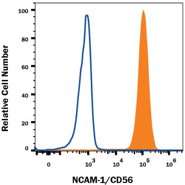
Detection of NCAM‑1/CD56 in Neuro‑2A Mouse Cell Line by Flow Cytometry.
Neuro-2A mouse neuroblastoma cell line was stained with Rat Anti-Mouse NCAM-1/CD56 Monoclonal Antibody (Catalog # MAB7820, filled histogram) or isotype control antibody (Catalog # MAB006, open histogram), followed by Allophycocyanin-conjugated Anti-Rat IgG Secondary Antibody (Catalog # F0113). View our protocol for Staining Membrane-associated Proteins.Applications for Mouse NCAM-1/CD56 Antibody
Application
Recommended Usage
COMET
CyTOF-ready
Ready to be labeled using established conjugation methods. No BSA or other carrier proteins that could interfere with conjugation.
Flow Cytometry
0.25 µg/106 cells
Sample: Neuro‑2A mouse neuroblastoma cell line
Sample: Neuro‑2A mouse neuroblastoma cell line
Immunohistochemistry
3-25 µg/mL
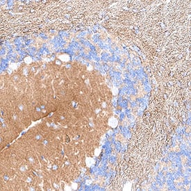
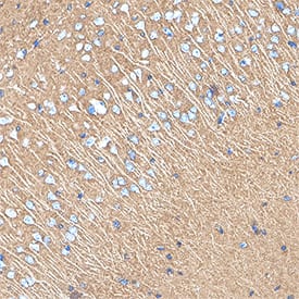
Sample: Immersion fixed paraffin-embedded sections of mouse cerebellum and mouse cortex
Sample: Immersion fixed paraffin-embedded sections of mouse cerebellum and mouse cortex
Multiplex Immunofluorescence
15 µg/mL
Sample: Immersion fixed paraffin-embedded sections of mouse cerebellum
Sample: Immersion fixed paraffin-embedded sections of mouse cerebellum
Western Blot
0.1 µg/mL
Sample: Neuro‑2A mouse neuroblastoma cell line
Sample: Neuro‑2A mouse neuroblastoma cell line
Formulation, Preparation, and Storage
Purification
Protein A or G purified from hybridoma culture supernatant
Reconstitution
Sterile PBS to a final concentration of 0.5 mg/mL. For liquid material, refer to CoA for concentration.
Formulation
Lyophilized from a 0.2 μm filtered solution in PBS with Trehalose. See Certificate of Analysis for details.
*Small pack size (-SP) is supplied either lyophilized or as a 0.2 µm filtered solution in PBS.
*Small pack size (-SP) is supplied either lyophilized or as a 0.2 µm filtered solution in PBS.
Shipping
Lyophilized product is shipped at ambient temperature. Liquid small pack size (-SP) is shipped with polar packs. Upon receipt, store immediately at the temperature recommended below.
Stability & Storage
Use a manual defrost freezer and avoid repeated freeze-thaw cycles.
- 12 months from date of receipt, -20 to -70 °C as supplied.
- 1 month, 2 to 8 °C under sterile conditions after reconstitution.
- 6 months, -20 to -70 °C under sterile conditions after reconstitution.
Background: NCAM-1/CD56
NCAM-1 (Neural adhesion molecule-1; also CD56) is a 120-190 kDa glycoprotein member of the Ig Superfamily. It is expressed on multiple cell types, both in the embryo and adult. Here, it serves as both an adhesion molecule and a receptor for multiple ligands, including as FGFR, PDGF, GDNF and agrin. On the cell surface, it is a cis-oriented homodimer that can form homodimers in-trans with other cis-homodimers. In the embryo, NCAM-1 is polysialylated (PolySia), and shows a MW of 200-220 kDa in SDS-PAGE. This polysialylation reduces the ability of NCAM-1 to dimerize. Mature mouse NCAM-1 is a 1096 amino acid (aa) type I transmembrane (TM) protein (aa 20-1115). It possesses a 692 aa extracellular region (aa 20-711) and a 386 aa cytoplasmic domain. The extracellular region contains five consecutive C2-type Ig-like domains (aa 20-492) followed by two FN type-III domains (aa 497-692). Multiple splice variants exist. There is a 140 kDa TM variant that shows a deletion of aa 810-1076, and a 120 kDa variant that is GPI-linked and shows a 24 aa substitution for aa 702-1115. A third potential variant contains a five aa substitution for aa 601-1115. Over aa 20-711, mouse NCAM-1 shares 99% and 95% aa identity with rat and human NCAM-1, respectively.
Long Name
Neural Cell Adhesion Molecule
Alternate Names
CD56, NCAM1
Gene Symbol
NCAM1
UniProt
Additional NCAM-1/CD56 Products
Product Documents for Mouse NCAM-1/CD56 Antibody
Product Specific Notices for Mouse NCAM-1/CD56 Antibody
For research use only
Loading...
Loading...
Loading...
Loading...
Loading...
Loading...