
Mouse Ki67/MKI67 Antibody
R&D Systems, part of Bio-Techne | Catalog # AF7649


Scientific images may be resized, cropped, or adjusted for brightness or contrast for presentation purposes. These updates are limited to presentation and do not affect the underlying scientific interpretation of the data. Where available, additional source data may be provided upon request.
Conjugate
Catalog #
Key Product Details
Species Reactivity
Validated:
Mouse
Cited:
Human, Mouse, Transgenic Mouse
Applications
Validated:
Immunocytochemistry
Cited:
Immunohistochemistry, Immunohistochemistry-Paraffin, Immunocytochemistry
Label
Unconjugated
Antibody Source
Polyclonal Sheep IgG
Product Specifications
Immunogen
E. coli-derived recombinant mouse Ki67/MKI67
Asn3053-Ser3177
Accession # NP_001074586
Asn3053-Ser3177
Accession # NP_001074586
Specificity
Detects mouse Ki67/MKI67 in direct ELISAs. In direct ELISAs, less than 5% cross-reactivity with recombinant human Ki67/MKI67 is observed.
Clonality
Polyclonal
Host
Sheep
Isotype
IgG
Scientific Data Images for Mouse Ki67/MKI67 Antibody
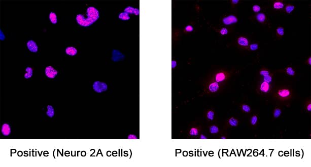
Detection of Ki67/MKI67 in RAW264.7cells and Neuro2A cells.
Ki67/MKI67 was detected in immersion fixed RAW 264.7 mouse monocyte/macrophage cell line (Positive) and Neuro-2A mouse neuroblastoma cell line (Positive) using Sheep Anti-Mouse Ki67/MKI67 Antigen Affinity-purified Polyclonal Antibody (Catalog # AF7649) at 1.7 µg/ml for 3 hours at room temperature. Cells were stained using the NorthernLights™ 557-conjugated Anti-Sheep IgG Secondary Antibody (red; Catalog # NL010) and counterstained with DAPI (blue). Specific staining was localized to the nucleus. View our protocol for Fluorescent ICC Staining of Cells on Coverslips.
Ki67/MKI67 in NIH‑3T3 Mouse Cell Line.
Ki67/MKI67 was detected in immersion fixed NIH-3T3 mouse embryonic fibroblast cell line using Sheep Anti-Mouse Ki67/MKI67 Antigen Affinity-purified Polyclonal Antibody (Catalog # AF7649) at 10 µg/mL for 3 hours at room temperature. Cells were stained using the NorthernLights™ 557-conjugated Anti-Sheep IgG Secondary Antibody (red, upper panel; Catalog # NL010) and counterstained with DAPI (blue, lower panel). Specific staining was localized to nuclei and nucleoli. View our protocol for Fluorescent ICC Staining of Cells on Coverslips.
Detection of Ki67/MKI67 by Immunohistochemistry
Representative images of pERK and Ki67 staining in uninjured mice 96 h after FGF7p. (a–h) Co‐IF for pERK (green) and Ki67 (red) in vehicle treated (Veh) (a–d) and FGF7p‐treated mice (FGF7p) (e–h). (a–d) Vehicle‐treated mice had no pERK+ or Ki67+ urothelial cells in the green channel (a), the red channel (b), merged images without DAPI (c) or merged images with DAPI (d). E‐H. FGF7p‐treated mice had subsets of urothelial cells that were pERK+ on the green channel (e, arrowhead) and Ki67+ cells some of which appeared to also express pERK (f, arrowhead) and others that did not (f, concave arrowhead). Merged images without DAPI (g) and with DAPI (h) confirmed presence of pERK+/Ki67+ cells (arrowheads) and others that were only Ki67+ (concave arrowheads). Scale bars (a–h) = 50 µm; Dashed line = Urothelial border with stroma; L, lumen Image collected and cropped by CiteAb from the following open publication (https://pubmed.ncbi.nlm.nih.gov/35388988), licensed under a CC-BY license. Not internally tested by R&D Systems.Applications for Mouse Ki67/MKI67 Antibody
Application
Recommended Usage
Immunocytochemistry
1-15 µg/mL
Sample: Immersion fixed NIH‑3T3 mouse embryonic fibroblast cell line, RAW264.7 mouse monocyte/macrophage cell line, and Neuro-2A mouse neurosblastoma cell line
Sample: Immersion fixed NIH‑3T3 mouse embryonic fibroblast cell line, RAW264.7 mouse monocyte/macrophage cell line, and Neuro-2A mouse neurosblastoma cell line
Reviewed Applications
Read 2 reviews rated 5 using AF7649 in the following applications:
Formulation, Preparation, and Storage
Purification
Antigen Affinity-purified
Reconstitution
Sterile PBS to a final concentration of 0.2 mg/mL. For liquid material, refer to CoA for concentration.
Formulation
Lyophilized from a 0.2 μm filtered solution in PBS with Trehalose. See Certificate of Analysis for details.
*Small pack size (-SP) is supplied either lyophilized or as a 0.2 µm filtered solution in PBS.
*Small pack size (-SP) is supplied either lyophilized or as a 0.2 µm filtered solution in PBS.
Shipping
Lyophilized product is shipped at ambient temperature. Liquid small pack size (-SP) is shipped with polar packs. Upon receipt, store immediately at the temperature recommended below.
Stability & Storage
Use a manual defrost freezer and avoid repeated freeze-thaw cycles.
- 12 months from date of receipt, -20 to -70 °C as supplied.
- 1 month, 2 to 8 °C under sterile conditions after reconstitution.
- 6 months, -20 to -70 °C under sterile conditions after reconstitution.
Background: Ki67/MKI67
MKI67 (also Ki67 and TSG126) is a 350-370 kDa nuclear protein that belongs to a molecular group comprised of mitotic chromosome-associated proteins. Ki67 was originally recognized as an antigen associated with the monoclonal Ki67 antibody raised against Hodgkin's lymphoma nuclear material. Ki67 is contextually expressed, being potentially found in all cells that are not in the Go phase of the cell cycle. Thus, MKI67 qualifies as a cell proliferation marker. Functionally, Ki67 is known to interact with 160 kDa Hklp2, a protein that promotes centrosome separation and spindle bipolarity. It also directly interacts with NIFK, and apparently binds to UBF, thus playing a role in rRNA synthesis. Mouse MKI67 is 3177 amino acids (aa) in length. It contains one FHA domain (aa 8-101), followed by sixteen 120 aa repeats (aa 993-2872). There are two potential isoform variants. One isoform shows a 19 aa substitution for 1120-3177, while a second isoform contains a deletion of aa 1169-1409. Over aa 3053-3177, mouse Ki67 shares 46% and 74% aa sequence identity with the human and rat orthologs to Ki67, respectively.
Long Name
Antigen Identified by Monoclonal Antibody Ki67
Alternate Names
Ki-67, KIA, MIB-1, MKI67, PPP1R105, TSG126
Gene Symbol
MKI67
UniProt
Additional Ki67/MKI67 Products
Product Documents for Mouse Ki67/MKI67 Antibody
Product Specific Notices for Mouse Ki67/MKI67 Antibody
For research use only
Loading...
Loading...
Loading...
Loading...
Loading...