
Mouse IFN-gamma Antibody
R&D Systems, part of Bio-Techne | Catalog # MAB4851


Scientific images may be resized, cropped, or adjusted for brightness or contrast for presentation purposes. These updates are limited to presentation and do not affect the underlying scientific interpretation of the data. Where available, additional source data may be provided upon request.
Conjugate
Catalog #
Key Product Details
Species Reactivity
Validated:
Mouse
Cited:
Mouse, Transgenic Mouse
Applications
Validated:
Neutralization, Immunocytochemistry
Cited:
Neutralization, Bioassay, In vivo assay
Label
Unconjugated
Antibody Source
Monoclonal Hamster IgG Clone # H22
Product Specifications
Immunogen
E. coli-derived recombinant mouse IFN-gamma
Accession # P01580
Accession # P01580
Specificity
Detects mouse IFN-gamma in direct ELISAs. Does not cross-react with rat, cotton rat, human, rhesus macaque, porcine, feline, or canine IFN-gamma.
Clonality
Monoclonal
Host
Hamster
Isotype
IgG
Endotoxin Level
<0.10 EU per 1 μg of the antibody by the LAL method.
Scientific Data Images for Mouse IFN-gamma Antibody
IFN‑ gamma Inhibition of EMCV-induced Cytopathy and Neutralization by Mouse IFN‑ gamma Antibody.
Recombinant Mouse IFN-gamma (Catalog # 485-MI) reduces the Encephalomyocarditis Virus (EMCV)-induced cytopathy in the L-929 mouse fibroblast cell line in a dose-dependent manner (orange line). Inhibition of EMCV activity elicited by Recombinant Mouse IFN-gamma (2.5 ng/mL) is neutralized (green line) by increasing concentrations of Hamster Anti-Mouse IFN-gamma Monoclonal Antibody (Catalog # MAB4851). The ND50 is typically 4-20 ng/mL.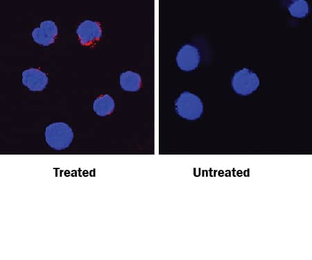
IFN‑ gamma in Mouse Splenocytes.
IFN‑ gamma was detected in immersion fixed mouse splenocytes stimulated with calcium ionomycin and PMA (left panel) or unstimulated (right panel) using Hamster Anti-Mouse IFN‑ gamma Monoclonal Antibody (Catalog # MAB4851) at 15 µg/mL for 3 hours at room temperature. Cells were stained using an anti-hamster IgG secondary antibody (red) and counterstained with DAPI (blue). Specific staining was localized to cytoplasm. View our protocol for Fluorescent ICC Staining of Non-adherent Cells.
Detection of IFN-gamma by Flow Cytometry
Dependency on FOXP3 in IFN gamma-mediated PD-L1 expression. A, Expression of FOXP3 and PD-L1 in bladder cancer HT1376, T24, and SW780 cell lines without and with 100 ng/mL IFN gamma stimulation by flow cytometry. The mean with SD of at least three independent experiments, P values indicated. *, P < 0.05; **, P < 0.01; ***, P < 0.001; ****, P < 0.0001. B, Expression of FOXP3 and PD-L1 in HT1376 CT and HT1376 FOXP3 KO bladder cancer cell lines without and with IFN gamma stimulation by qPCR. The mean of triplicates and representative of three independent experiments, P values indicated. C, Expression of FOXP3 and PD-L1 in HT1376 CT and HT1376 FOXP3 KO lines without and with IFN gamma stimulation by Western blotting. Sizes in kD as indicated. Sizes of FOXP3 delta3 and FOXP3 depicted by overexpression in 293T cells. Full Western blots shown in Supplementary Fig. S1. D, Flow cytometry of FOXP3 and PD-L1 expression in HT1376 CT and HT1376 FOXP3 KO lines without and with IFN gamma stimulation. Representative of four independent experiments. E, Expression of FOXP3 and PD-L1 in rescue of a clonal HT1376 FOXP3 KO line by FOXP3 delta3 and FOXP3 overexpression. Representative of three independent experiments. F, Expression of FOXP3 and PD-L1 in HT1376 FOXP3 KO lines with reconstitution of FOXP3 delta3 and FOXP3 by Western blotting. Sizes in kD as indicated. Full Western blots shown in Supplementary Fig. S1. G, Flow cytometry of PD-L1 in HT1376 CT and HT1376 FOXP3 KO lines stimulated by IFN gamma without and with the presence of JAK1/2 inhibitor ruxolitinib (20 μmol/L), P values indicated. H, Expression of phosphorylated STAT1 (P-STAT1) in HT1376 CT and HT1376 FOXP3 KO lines stimulated by IFN gamma without and with the presence of JAK1/2 inhibitor ruxolitinib by Western blot. Full Western blots shown in Supplementary Fig. S1. Image collected and cropped by CiteAb from the following open publication (https://pubmed.ncbi.nlm.nih.gov/39099201), licensed under a CC-BY license. Not internally tested by R&D Systems.Applications for Mouse IFN-gamma Antibody
Application
Recommended Usage
Immunocytochemistry
8-25 µg/mL
Sample: Immersion fixed mouse splenocytes stimulated with calcium ionomycin and PMA
Sample: Immersion fixed mouse splenocytes stimulated with calcium ionomycin and PMA
Neutralization
Measured by its ability to neutralize IFN‑ gamma inhibition of EMCV-induced cytopathy in the L‑929 mouse fibroblast cell line. Schreiber, R. et al. (1985) J. Immunol. 134:1609; Vogel, S. and M. Hogan (1995) in Current Protocols in Immunology. Ciocio, R. (ed); John Wiley & Sons, Inc. p. 6. 9. 1. The Neutralization Dose (ND50) is typically 4-20 ng/mL in the presence of 2.5 ng/mL Recombinant Mouse IFN‑ gamma.
Formulation, Preparation, and Storage
Purification
Protein A or G purified from hybridoma culture supernatant
Reconstitution
Reconstitute at 0.5 mg/mL in sterile PBS. For liquid material, refer to CoA for concentration.
Formulation
Lyophilized from a 0.2 μm filtered solution in PBS with Trehalose. *Small pack size (SP) is supplied either lyophilized or as a 0.2 µm filtered solution in PBS.
Shipping
Lyophilized product is shipped at ambient temperature. Liquid small pack size (-SP) is shipped with polar packs. Upon receipt, store immediately at the temperature recommended below.
Stability & Storage
Use a manual defrost freezer and avoid repeated freeze-thaw cycles.
- 12 months from date of receipt, -20 to -70 °C as supplied.
- 1 month, 2 to 8 °C under sterile conditions after reconstitution.
- 6 months, -20 to -70 °C under sterile conditions after reconstitution.
Background: IFN-gamma
Long Name
Interferon gamma
Alternate Names
IFG, IFI, IFNG, IFNgamma
Entrez Gene IDs
Gene Symbol
IFNG
UniProt
Additional IFN-gamma Products
Product Documents for Mouse IFN-gamma Antibody
Product Specific Notices for Mouse IFN-gamma Antibody
For research use only
Loading...
Loading...
Loading...
Loading...
Loading...