
Mouse Galectin-3 Biotinylated Antibody
R&D Systems, part of Bio-Techne | Catalog # BAF1197

Scientific images may be resized, cropped, or adjusted for brightness or contrast for presentation purposes. These updates are limited to presentation and do not affect the underlying scientific interpretation of the data. Where available, additional source data may be provided upon request.
Conjugate
Catalog #
Key Product Details
Species Reactivity
Mouse
Applications
Immunohistochemistry, Western Blot, ELISA Detection (Matched Antibody Pair)
Label
Biotin
Antibody Source
Polyclonal Goat IgG
Product Specifications
Immunogen
E. coli-derived recombinant mouse Galectin‑3
Ala2-Ile264
Accession # NP_034835
Ala2-Ile264
Accession # NP_034835
Specificity
Detects mouse Galectin-3 in ELISAs and Western blots. In sandwich ELISAs, approximately 10% cross‑reactivity with recombinant human (rh) Galectin-3 is observed and less than 0.05% cross-reactivity with recombinant mouse (rm) Galectin-1, rhGalectin-2, and rhGalectin-4 is observed.
Clonality
Polyclonal
Host
Goat
Isotype
IgG
Scientific Data Images for Mouse Galectin-3 Biotinylated Antibody
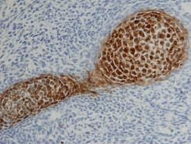
Galectin-3 in Mouse Cartilage.
Galectin-3 was detected in immersion fixed frozen sections of mouse cartilage using 15 µg/mL Goat Anti-Mouse Galectin-3 Biotinylated Antigen Affinity-purified Polyclonal Antibody (Catalog # BAF1197) overnight at 4 °C. Tissue was stained with the Anti-Goat HRP-DAB Cell & Tissue Staining Kit (brown; Catalog # CTS008) and counterstained with hematoxylin (blue). View our protocol for Chromogenic IHC Staining of Frozen Tissue Sections.
Mouse Galectin-3 ELISA Standard Curve
Recombinant Mouse Galectin-3 (Catalog # 1197-GA) was serially diluted and captured by Rat Anti-Human/Mouse/Rat Galectin-3 Monoclonal Antibody (Catalog # MAB1197) coated on a Clear Polystyrene Microplate (Catalog # DY990). Goat Anti-Human/Mouse/Rat Galectin-3 Antigen Affinity-purified Polyclonal Antibody (Catalog # AF1197) was biotinylated and incubated with the protein captured on the plate. Detection of the standard curve was achieved by incubating Streptavidin-HRP (Catalog # DY998)Applications for Mouse Galectin-3 Biotinylated Antibody
Application
Recommended Usage
Immunohistochemistry
5-15 µg/mL
Sample: Immersion fixed frozen sections of mouse cartilage
Sample: Immersion fixed frozen sections of mouse cartilage
Western Blot
0.1 µg/mL
Sample: Recombinant Mouse Galectin-3 (Catalog # 1197-GA)
Sample: Recombinant Mouse Galectin-3 (Catalog # 1197-GA)
Mouse Galectin-3 Sandwich Immunoassay
Please Note: Optimal dilutions of this antibody should be experimentally determined.
Formulation, Preparation, and Storage
Purification
Antigen Affinity-purified
Reconstitution
Reconstitute at 0.2 mg/mL in sterile PBS.
Formulation
Lyophilized from a 0.2 μm filtered solution in PBS with BSA as a carrier protein.
Shipping
The product is shipped at ambient temperature. Upon receipt, store it immediately at the temperature recommended below.
Stability & Storage
Use a manual defrost freezer and avoid repeated freeze-thaw cycles.
- 12 months from date of receipt, -20 to -70 °C as supplied.
- 1 month, 2 to 8 °C under sterile conditions after reconstitution.
- 6 months, -20 to -70 °C under sterile conditions after reconstitution.
Background: Galectin-3
The galectins constitute a large family of carbohydrate-binding proteins with specificity for N-acetyl-lactosamine-containing glycoproteins. At least 14 mammalian galectins, which share structural similarities in their carbohydrate recognition domains (CRD), have been identified. The galectins have been classified into the prototype galectins (-1, -2, -5, -7, -10, -11, -13, -14), which contain one CRD and exist either as a monomer or a noncovalent homodimer; the chimera galectins (Galectin-3) containing one CRD linked to a nonlectin domain; and the tandem-repeat galectins (-4, -6, -8, -9, -12) consisting of two CRDs joined by a linker peptide. Galectins lack a classical signal peptide and can be localized to the cytosolic compartments where they have intracellular functions. However, via one or more as yet unidentified non-classical secretory pathways, galectins can also be secreted to function extracellularly. Individual members of the galectin family have different tissue distribution profiles and exhibit subtle differences in their carbohydrate-binding specificities. Each family member may preferentially bind to a unique subset of cell-surface glycoproteins (1-4). Galectin-3, also known as Mac-2, L29, CBP35, and epsilonBP, is a chimera galectin that has a tendency to dimerize. Besides the soluble protein, alternatively spliced forms of chicken Galectin-3 containing a transmembrane-spanning domain and a leucine zipper motif have been reported. Galectin-3 is expressed in tumor cells, macrophages, activated T cells, osteoclasts, epithelial cells, and fibroblasts. It binds various matrix glycoproteins including laminin, fibronectin, LAMPS, 90K/Mac-2BP, MP20, and CEA. Galectin-3 promotes cell growth and proliferation for many cell types. Galectin-3 acts intracellularly to prevent apoptosis. Depending on the cell types, Galectin-3 exhibits pro- or anti-adhesive properties. Galectin-3 has proinflammatory activities in vitro and in vivo. It induces pro-inflammatory and inhibits Th2 type cytokine production. Galectin-3 chemoattracts monocytes and macrophages. It activates and degranulates basophils and mast cells. Elevated circulating levels of Galectin-3 has been show to correlate with the malignant potential of several types of cancer, suggesting that Galectin-3 is also involved in tumor growth and metastasis. Human and mouse Galectin-3 shares approximately 80% amino acid sequence similarity (1-5).
References
- Rabinovich, A. et al. (2002) Trends in Immunol. 23:313.
- Rabinovich, A. et al. (2002) J. Leukocyte Biology 71:741.
- Hughes, R.C. (2001) Biochimie 83:667.
- R&D Systems Cytokine Bulletin; Summer 2002.
- Gorski, J.P. et al. (2002) J. Biol. Chem. 277:18840.
Alternate Names
AGE-R3, CBP35, GAL3, Galectin3, L29, LGALS3, Mac-2
Gene Symbol
LGALS3
UniProt
Additional Galectin-3 Products
Product Documents for Mouse Galectin-3 Biotinylated Antibody
Product Specific Notices for Mouse Galectin-3 Biotinylated Antibody
For research use only
Loading...
Loading...
Loading...
Loading...
Loading...