
Mouse Fetuin A/AHSG Antibody
R&D Systems, part of Bio-Techne | Catalog # AF1563

Scientific images may be resized, cropped, or adjusted for brightness or contrast for presentation purposes. These updates are limited to presentation and do not affect the underlying scientific interpretation of the data. Where available, additional source data may be provided upon request.
Conjugate
Catalog #
Key Product Details
Species Reactivity
Validated:
Mouse
Cited:
Human, Mouse
Applications
Validated:
Western Blot
Cited:
Immunohistochemistry-Paraffin, Western Blot, Immunoprecipitation
Label
Unconjugated
Antibody Source
Polyclonal Goat IgG
Product Specifications
Immunogen
Mouse myeloma cell line NS0-derived recombinant mouse Fetuin A/AHSG (R&D Systems, Catalog # 1563-PI)
Ala19-Ile345
Accession # NP_038493
Ala19-Ile345
Accession # NP_038493
Specificity
Detects mouse Fetuin A/AHSG in direct ELISAs and Western blots. In these formats, approximately 5% cross-reactivity with recombinant human Fetuin A/AHSG is observed.
Clonality
Polyclonal
Host
Goat
Isotype
IgG
Scientific Data Images for Mouse Fetuin A/AHSG Antibody
Detection of Fetuin A/AHSG by Western Blot
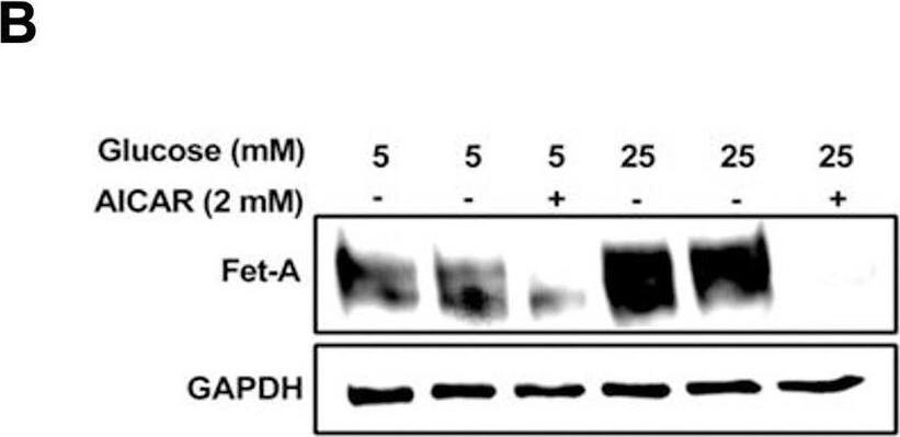
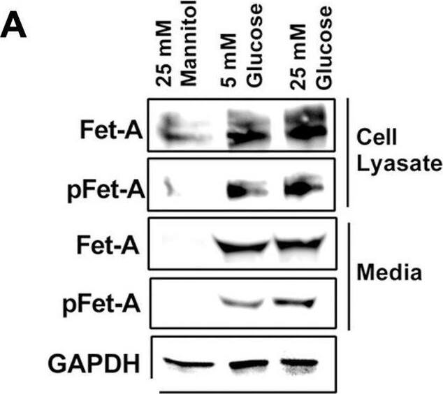
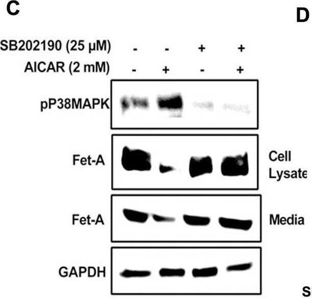
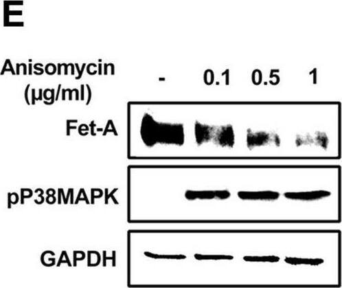
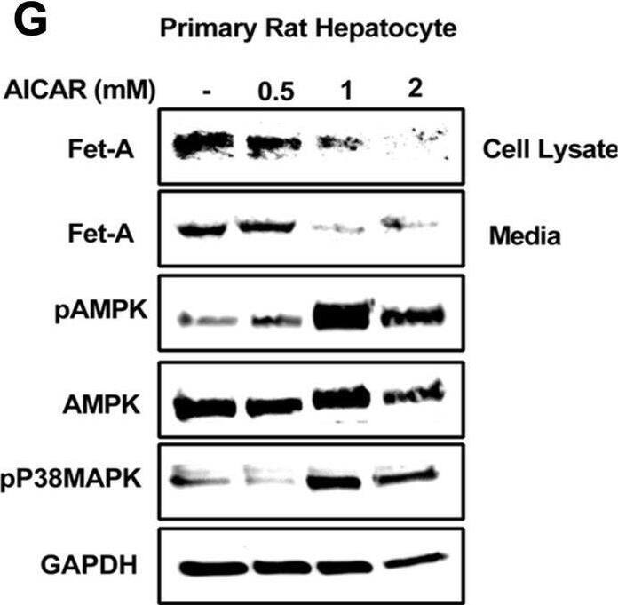
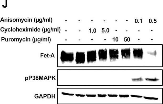
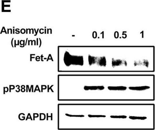
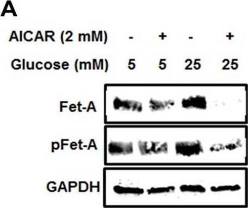
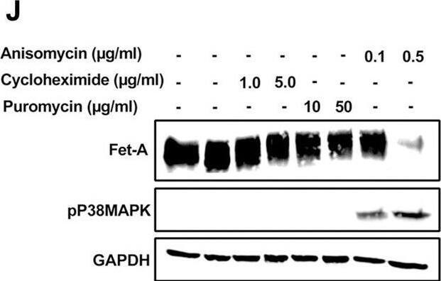
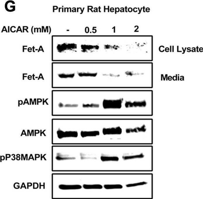
Activation of AMPK downregulates high glucose-induced Fet-A expression in HepG2 cells.[A] HepG2 cells were incubated in a media containing either mannitol, low glucose or high glucose for 12 hours, and cell lysates/media were subjected to immunoblotting for Fet-A and pFet-A (n = 3). [B] HepG2 cells were incubated with either low- or high-glucose in the absence or presence of AICAR for 12 h and cell lysates were analyzed by Western blotting. The blots were analyzed with antibodies against Fet-A (n = 4). [C] HepG2 cells were incubated with increasing concentrations (0.5, 1, 2 mM) of AICAR for 12 h. Cell lysate or media were analyzed by Western blotting for indicated proteins (n = 3) and [D] level of Fet-A and pFet-A in media, as a ratio of GAPDH were expressed. [E] HepG2 cells were incubated in low or high glucose in the absence or presence of AICAR/metformin for 12 hours, and media was used to detect Fet-A by ELISA technique (n = 4). [F] Real-time gene expression analysis was carried out for Fet-A after AICAR and metformin treatment (n = 4). [G] HepG2 cells were incubated with AICAR/metformin in the presence or absence of Compound C, an AMPK inhibitor. Cell lysates were immunoblotted for pAMPK, Fet-A as well as p-Fet-A and [H] Fet-A levels, as a ratio to GAPDH are depicted (n = 4). Data are shown as Means ± SEM. P values were determined by one-way ANOVA followed by Tukey’s multiple comparison tests (C-F). Data are representative of at least three independent experiments performed in replicates. * Indicates p < 0.05. Image collected and cropped by CiteAb from the following open publication (https://pubmed.ncbi.nlm.nih.gov/35522655), licensed under a CC-BY license. Not internally tested by R&D Systems.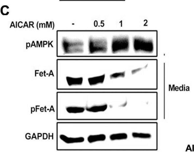
Detection of Fetuin A/AHSG by Western Blot
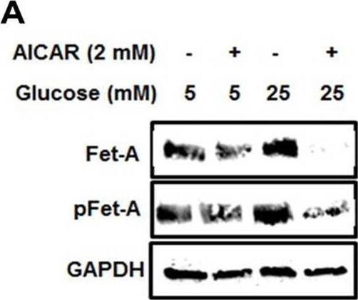
Activation of AMPK downregulates high glucose-induced Fet-A expression in HepG2 cells.[A] HepG2 cells were incubated in a media containing either mannitol, low glucose or high glucose for 12 hours, and cell lysates/media were subjected to immunoblotting for Fet-A and pFet-A (n = 3). [B] HepG2 cells were incubated with either low- or high-glucose in the absence or presence of AICAR for 12 h and cell lysates were analyzed by Western blotting. The blots were analyzed with antibodies against Fet-A (n = 4). [C] HepG2 cells were incubated with increasing concentrations (0.5, 1, 2 mM) of AICAR for 12 h. Cell lysate or media were analyzed by Western blotting for indicated proteins (n = 3) and [D] level of Fet-A and pFet-A in media, as a ratio of GAPDH were expressed. [E] HepG2 cells were incubated in low or high glucose in the absence or presence of AICAR/metformin for 12 hours, and media was used to detect Fet-A by ELISA technique (n = 4). [F] Real-time gene expression analysis was carried out for Fet-A after AICAR and metformin treatment (n = 4). [G] HepG2 cells were incubated with AICAR/metformin in the presence or absence of Compound C, an AMPK inhibitor. Cell lysates were immunoblotted for pAMPK, Fet-A as well as p-Fet-A and [H] Fet-A levels, as a ratio to GAPDH are depicted (n = 4). Data are shown as Means ± SEM. P values were determined by one-way ANOVA followed by Tukey’s multiple comparison tests (C-F). Data are representative of at least three independent experiments performed in replicates. * Indicates p < 0.05. Image collected and cropped by CiteAb from the following open publication (https://pubmed.ncbi.nlm.nih.gov/35522655), licensed under a CC-BY license. Not internally tested by R&D Systems.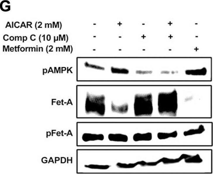
Detection of Fetuin A/AHSG by Western Blot
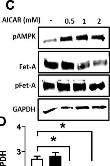
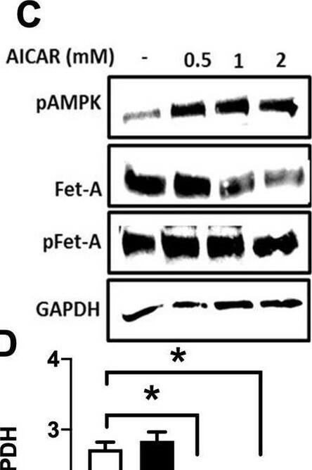
Activation of AMPK downregulates high glucose-induced Fet-A expression in HepG2 cells.[A] HepG2 cells were incubated in a media containing either mannitol, low glucose or high glucose for 12 hours, and cell lysates/media were subjected to immunoblotting for Fet-A and pFet-A (n = 3). [B] HepG2 cells were incubated with either low- or high-glucose in the absence or presence of AICAR for 12 h and cell lysates were analyzed by Western blotting. The blots were analyzed with antibodies against Fet-A (n = 4). [C] HepG2 cells were incubated with increasing concentrations (0.5, 1, 2 mM) of AICAR for 12 h. Cell lysate or media were analyzed by Western blotting for indicated proteins (n = 3) and [D] level of Fet-A and pFet-A in media, as a ratio of GAPDH were expressed. [E] HepG2 cells were incubated in low or high glucose in the absence or presence of AICAR/metformin for 12 hours, and media was used to detect Fet-A by ELISA technique (n = 4). [F] Real-time gene expression analysis was carried out for Fet-A after AICAR and metformin treatment (n = 4). [G] HepG2 cells were incubated with AICAR/metformin in the presence or absence of Compound C, an AMPK inhibitor. Cell lysates were immunoblotted for pAMPK, Fet-A as well as p-Fet-A and [H] Fet-A levels, as a ratio to GAPDH are depicted (n = 4). Data are shown as Means ± SEM. P values were determined by one-way ANOVA followed by Tukey’s multiple comparison tests (C-F). Data are representative of at least three independent experiments performed in replicates. * Indicates p < 0.05. Image collected and cropped by CiteAb from the following open publication (https://pubmed.ncbi.nlm.nih.gov/35522655), licensed under a CC-BY license. Not internally tested by R&D Systems.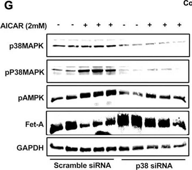
Applications for Mouse Fetuin A/AHSG Antibody
Application
Recommended Usage
Western Blot
0.1 µg/mL
Sample: Recombinant Mouse Fetuin A/AHSG (Catalog # 1563-PI)
Sample: Recombinant Mouse Fetuin A/AHSG (Catalog # 1563-PI)
Reviewed Applications
Read 1 review rated 5 using AF1563 in the following applications:
Formulation, Preparation, and Storage
Purification
Antigen Affinity-purified
Reconstitution
Reconstitute at 0.2 mg/mL in sterile PBS. For liquid material, refer to CoA for concentration.
Formulation
Lyophilized from a 0.2 μm filtered solution in PBS with Trehalose. *Small pack size (SP) is supplied either lyophilized or as a 0.2 µm filtered solution in PBS.
Shipping
Lyophilized product is shipped at ambient temperature. Liquid small pack size (-SP) is shipped with polar packs. Upon receipt, store immediately at the temperature recommended below.
Stability & Storage
Use a manual defrost freezer and avoid repeated freeze-thaw cycles.
- 12 months from date of receipt, -20 to -70 °C as supplied.
- 1 month, 2 to 8 °C under sterile conditions after reconstitution.
- 6 months, -20 to -70 °C under sterile conditions after reconstitution.
Background: Fetuin A/AHSG
Mouse Fetuin A, also known as alpha2-Heremans-Schmid glycoprotein, is encoded by the AHSG gene. It has been also called "countertrypsin" because of its ability to inhibit trypsin (1). It is a major plasma protein and a member of the cystatin superfamily of protease inhibitors (2, 3). It is expressed by hepatocytes, the principal cell source, and by monocyte/macrophages (4). The major form of plasma Fetuin A corresponds to two disulfide bond-linked chains derived from the single chain (5). Fetuin-A has a number of functions. It is a negative acute-phase protein with normal circulating levels in adults (300-600 μg/mL), which fall significantly (30-50%) during injury and infection (5). It enhances entry of cationic inhibitors into macrophages (6). It inhibits both insulin receptor autophosphorylation and undesirable calcification (7, 8).
References
- Yamamoto, K. and H. Sinohara (1993) J. Biol. Chem. 268:17750.
- Kellemann, J. et al. (1989) J. Biol. Chem. 264:14121.
- Dziegielewska, K.M. et al. (1990) J. Biol. Chem. 265:4354.
- Dziegielewska, K.M. et al. (1996) Histochem. Cell Biol. 106:319.
- Gejyo, F. and K. Schmid (1981) Biochim. Biophys. Acta 671:78.
- Wang, H. et al. (1998) Proc. Natl. Acad. Sci. USA 95:14429.
- Mathews, S.T. et al. (2000) Mol. Cell Endocrinol. 164:87.
- Schäfer, C. et al. (2003) J. Clin. Invest. 112:357.
Long Name
alpha-2-HS Glycoprotein
Alternate Names
AHSG, alpha-2-HS-glycoprotein
Gene Symbol
AHSG
UniProt
Additional Fetuin A/AHSG Products
Product Documents for Mouse Fetuin A/AHSG Antibody
Product Specific Notices for Mouse Fetuin A/AHSG Antibody
For research use only
Loading...
Loading...
Loading...
Loading...
Loading...