
Mouse EphA2 Antibody
R&D Systems, part of Bio-Techne | Catalog # AF639

Scientific images may be resized, cropped, or adjusted for brightness or contrast for presentation purposes. These updates are limited to presentation and do not affect the underlying scientific interpretation of the data. Where available, additional source data may be provided upon request.
Conjugate
Catalog #
Key Product Details
Species Reactivity
Validated:
Mouse
Cited:
Human, Mouse, Transgenic Mouse
Applications
Validated:
Immunohistochemistry, Western Blot
Cited:
Immunohistochemistry, Immunohistochemistry-Paraffin, Immunohistochemistry-Frozen, Western Blot
Label
Unconjugated
Antibody Source
Polyclonal Goat IgG
Product Specifications
Immunogen
Mouse myeloma cell line NS0-derived recombinant mouse EphA2
Ala22-Ala535
Accession # AAA82113
Ala22-Ala535
Accession # AAA82113
Specificity
Detects mouse EphA2 in direct ELISAs and Western blots. In direct ELISAs, approximately 10% cross-reactivity with recombinant human (rh) EphA2 is observed, and less than 1% cross‑reactivity with rhEphA1, recombinant mouse (rm) EphA3, rmEphA4, rmEphA6, rmEphA7, rmEphA8, and recombinant rat EphA5 is observed.
Clonality
Polyclonal
Host
Goat
Isotype
IgG
Scientific Data Images for Mouse EphA2 Antibody
Detection of EphA2 by Western Blot
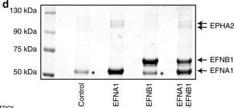
EPHA2 is critical for EFN ligand responses in mESCs.a Average protein copy number per cell determined for receptor kinases in mESCs, using quantitative whole-cell proteomics. Data are presented as mean ± SD (n = 3). b Workflow for quantification of EPH–EFN interactions in mESCs by EFN ligand affinity purification mass spectrometry. c Proof-of-principle identification of EPH–EFN interactions by EFN ligand affinity purification. EPHA2 levels were determined by immunoblotting. d Coomassie staining of EFNA1/B1 affinity purification from mESCs. EFNA1, EFNB1 and EPHA2 proteins are indicated. (*) = non-specific band. e Mass-spectrometry analysis of 75–130-kDa region of the Coomassie stained EFNA1/EFNB1 affinity purification shown in (d). Total spectral counts recovered for each EPH receptor family member are indicated. f EFNA1/B1 affinity purification from intact Epha2+/+ and pooled Epha2−/− mESCs. Phosphotyrosine (pTyr), EPHA2 and ERK1/2 levels were determined by immunoblotting. Note that the pTyr signal is specific for EPHA2, and is not detected in the absence of EFN ligand. g EPHA2 was immunoprecipitated from Epha2+/+ and Epha2−/− mESCs, and pTyr, EPHA2, EFNA1 and ERK1/2 levels determined by immunoblotting. hEpha2+/+ mESCs were stimulated with EFNA1-expressing Epha2−/− mESCs for 15 or 40 min. EPHA2 was immunoprecipitated, and pTyr and EPHA2 levels determined by immunoblotting. Source data are provided as a Source Data file. Image collected and cropped by CiteAb from the following open publication (https://pubmed.ncbi.nlm.nih.gov/32170114), licensed under a CC-BY license. Not internally tested by R&D Systems.Applications for Mouse EphA2 Antibody
Application
Recommended Usage
Immunohistochemistry
5-15 µg/mL
Sample: Immersion fixed frozen sections of mouse embryo (E11)
Sample: Immersion fixed frozen sections of mouse embryo (E11)
Western Blot
0.1 µg/mL
Sample: Recombinant Mouse EphA2 Fc Chimera (Catalog # 639-A2)
Sample: Recombinant Mouse EphA2 Fc Chimera (Catalog # 639-A2)
Formulation, Preparation, and Storage
Purification
Antigen Affinity-purified
Reconstitution
Reconstitute at 0.2 mg/mL in sterile PBS. For liquid material, refer to CoA for concentration.
Formulation
Lyophilized from a 0.2 μm filtered solution in PBS with Trehalose. *Small pack size (SP) is supplied either lyophilized or as a 0.2 µm filtered solution in PBS.
Shipping
Lyophilized product is shipped at ambient temperature. Liquid small pack size (-SP) is shipped with polar packs. Upon receipt, store immediately at the temperature recommended below.
Stability & Storage
Use a manual defrost freezer and avoid repeated freeze-thaw cycles.
- 12 months from date of receipt, -20 to -70 °C as supplied.
- 1 month, 2 to 8 °C under sterile conditions after reconstitution.
- 6 months, -20 to -70 °C under sterile conditions after reconstitution.
Background: EphA2
EphA2, also known as Eck, Myk2, and Sek2 (1), is a member of the Eph receptor family which binds members of the ephrin ligand family. There are two classes of receptors, designated A and B. Both the A and B class receptors have an extracellular region consisting of a globular domain, a cysteine-rich domain, and two fibronectin type III domains. This is followed by the transmembrane region and cytoplasmic region. The cytoplasmic region contains a juxtamembrane motif with two tyrosine residues, which are the major autophosphorylation sites, a kinase domain, and a conserved sterile alpha motif (SAM) in the carboxy tail which contains one conserved tyrosine residue. Activation of kinase activity occurs after ligand recognition and binding. EphA2 has been shown to bind ephrin-A3, ephrin-A1, ephrin-A5, ephrin-A4, and ephrin-A2 (2, 3). The extracellular domains of mouse and human EphA2 share greater than 92% amino acid identity. Only membrane-bound or Fc‑clustered ligands are capable of activating the receptor in vitro. While soluble monomeric ligands bind the receptor, they do not induce receptor autophosphorylation and activation (2). In vivo, the ligands and receptors display reciprocal expression (3). It has been found that nearly all receptors and ligands are expressed in developing and adult neural tissue (3). The Eph/ephrin families also appear to play a role in angiogenesis (3).
References
- Eph Nomenclature Committee [letter] (1997) Cell 90:403.
- Flanagan, J.G. and P. Vanderhaegen (1998) Annu. Rev. Neurosci. 21:309.
- Pasquale, E.B. (1997) Curr. Opin. Cell. Biol. 9:608.
Alternate Names
Eck, Myk2, Sek2
Gene Symbol
EPHA2
UniProt
Additional EphA2 Products
Product Documents for Mouse EphA2 Antibody
Product Specific Notices for Mouse EphA2 Antibody
For research use only
Loading...
Loading...
Loading...
Loading...
Loading...