
Mouse CHL-1/L1CAM-2 Antibody
R&D Systems, part of Bio-Techne | Catalog # AF2147

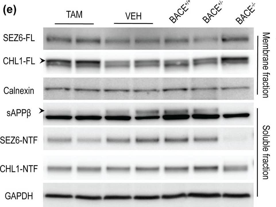

Scientific images may be resized, cropped, or adjusted for brightness or contrast for presentation purposes. These updates are limited to presentation and do not affect the underlying scientific interpretation of the data. Where available, additional source data may be provided upon request.
Conjugate
Catalog #
Key Product Details
Validated by
Biological Validation
Species Reactivity
Validated:
Mouse
Cited:
Human, Mouse, Rat, Transgenic Mouse
Applications
Validated:
Immunohistochemistry, Western Blot, Simple Western
Cited:
Immunohistochemistry, Immunohistochemistry-Frozen, Western Blot, Neutralization, Flow Cytometry, Immunocytochemistry, Immunoprecipitation, Cell Culture, Mass Spectrometry
Label
Unconjugated
Antibody Source
Polyclonal Goat IgG
Product Specifications
Immunogen
Mouse myeloma cell line NS0-derived recombinant mouse CHL‑1/L1CAM‑2
Ala25-Gln1043 (Leu227-Gln242 del, Ala243Ser)
Accession # BAC30699
Ala25-Gln1043 (Leu227-Gln242 del, Ala243Ser)
Accession # BAC30699
Specificity
Detects mouse CHL‑1/L1CAM‑2 in direct ELISAs and Western blots. In direct ELISAs and Western blots, approximately 40% cross-reactivity with recombinant human CHL-1 is observed.
Clonality
Polyclonal
Host
Goat
Isotype
IgG
Scientific Data Images for Mouse CHL-1/L1CAM-2 Antibody
Detection of Mouse CHL‑1/L1CAM‑2 by Western Blot.
Western blot shows lysates of mouse brain stem tissue. PVDF membrane was probed with 0.25 µg/mL of Goat Anti-Mouse CHL-1/L1CAM-2 Antigen Affinity-purified Polyclonal Antibody (Catalog # AF2147) followed by HRP-conjugated Anti-Goat IgG Secondary Antibody (Catalog # HAF019). A specific band was detected for CHL-1/L1CAM-2 at approximately 200 kDa (as indicated). This experiment was conducted under reducing conditions and using Immunoblot Buffer Group 1.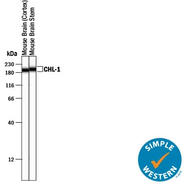
Detection of Mouse CHL‑1/L1CAM‑2 by Simple WesternTM.
Simple Western lane view shows lysates of mouse brain (cortex) tissue and mouse brain stem tissue, loaded at 0.2 mg/mL. A specific band was detected for CHL-1/L1CAM-2 at approximately 196-201 kDa (as indicated) using 2.5 µg/mL of Goat Anti-Mouse CHL-1/L1CAM-2 Antigen Affinity-purified Polyclonal Antibody (Catalog # AF2147) followed by 1:50 dilution of HRP-conjugated Anti-Goat IgG Secondary Antibody (Catalog # HAF109) . This experiment was conducted under reducing conditions and using the 12-230 kDa separation system.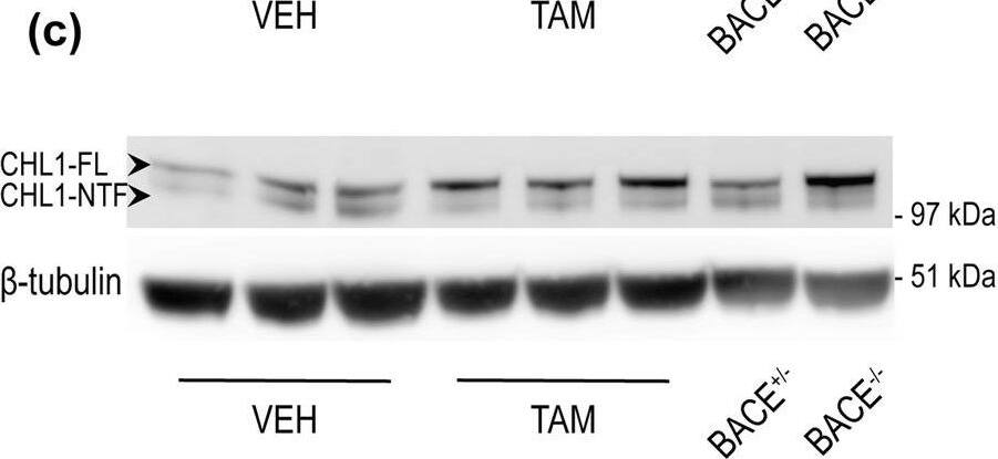
Detection of Mouse CHL-1/L1CAM-2 by Western Blot
BACE1-mediated processing of APP and CHL1 is reduced in cortex of aged BACE1 cKO mice following tamoxifen treatment. Cortex homogenates from TAM- or VEH-treated mice were resolved by SDS-PAGE for Western blot analysis of APP and CHL1 processing. Homogenates from aged-matched BACE+/− and BACE1−/− were also loaded as control samples. APP-FL, pC99 and pC89 were normalized to GAPDH (MAB374) while CHL1-FL and CHL1-NTF were normalized to beta-tubulin (JDR.3B8). Protein amount was normalized to protein levels in control mice injected with vehicle (set at 1). Representative blots of (a) APP-FL (C1/6.1), (b) APP-CTFs (C1/6.1) and (c) CHL1. (d) Densitometry analysis of protein expression. APP processing was reduced in TAM-treated mice as demonstrated by the accumulation of APP-FL (C1/6.1), and reduced levels of the betaCTFs pC99 and pC89. betaCTFs were clearly identified because missing in the BACE1−/− sample. CHL1-FL (AF2147) levels were increased and CHL1-NTF levels were significantly reduced. Furthermore, the CHL1-NTF/CHL1-FL ratio was significantly decreased in TAM-treated mice demonstrating reduced BACE1 processing (VEH n = 7; TAM n = 7). (e) Quantification of A betax-40 was performed by MSD immunoassay on cortex homogenates and expressed as pMol/g of cortex. The decrease of levels of A betax-40 in TAM-treated mice was comparable to the one observed in samples collected from young TAM-treated mice (~50% decrease) (VEH n = 7; TAM n = 7). Results were plotted as Mean ± SEM, *p < 0.05; **p < 0.005; ***p < 0.001; ****p < 0.0001; n.s. = not significant, Student’s t test. Image collected and cropped by CiteAb from the following publication (https://pubmed.ncbi.nlm.nih.gov/31882662), licensed under a CC-BY license. Not internally tested by R&D Systems.
Applications for Mouse CHL-1/L1CAM-2 Antibody
Application
Recommended Usage
Immunohistochemistry
5-15 µg/mL
Sample: Perfusion fixed frozen sections of mouse brain (cortex)
Sample: Perfusion fixed frozen sections of mouse brain (cortex)
Simple Western
2.5 µg/mL
Sample: Mouse brain (cortex) tissue and mouse brain stem tissue
Sample: Mouse brain (cortex) tissue and mouse brain stem tissue
Western Blot
0.25 µg/mL
Sample: Mouse brain stem tissue
Sample: Mouse brain stem tissue
Reviewed Applications
Read 4 reviews rated 4.8 using AF2147 in the following applications:
Formulation, Preparation, and Storage
Purification
Antigen Affinity-purified
Reconstitution
Reconstitute at 0.2 mg/mL in sterile PBS. For liquid material, refer to CoA for concentration.
Formulation
Lyophilized from a 0.2 μm filtered solution in PBS with Trehalose. *Small pack size (SP) is supplied either lyophilized or as a 0.2 µm filtered solution in PBS.
Shipping
Lyophilized product is shipped at ambient temperature. Liquid small pack size (-SP) is shipped with polar packs. Upon receipt, store immediately at the temperature recommended below.
Stability & Storage
Use a manual defrost freezer and avoid repeated freeze-thaw cycles.
- 12 months from date of receipt, -20 to -70 °C as supplied.
- 1 month, 2 to 8 °C under sterile conditions after reconstitution.
- 6 months, -20 to -70 °C under sterile conditions after reconstitution.
Background: CHL-1/L1CAM-2
Close homolog of L1 (CHL-1), also known as cell adhesion L1-like (CALL) and L1 cell adhesion molecule 2 (L1CAM-2), belongs to the L1 subfamily of immunoglobulin (Ig) superfamily cell adhesion molecules, which also includes L1, neurofascin and NgCAM-related cell adhesion molecule (NrCAM) (1‑3). These molecules are type I transmembrane proteins that have 6 Ig-like domains and 4‑5 fibronectin type III-like (FNIII) domains in their extracellular regions. They also share a highly conserved cytoplasmic region of approximately 110 amino acid (aa) residues containing an ankyrin-binding site. CHL-1 is expressed as a highly glycosylated 185 kDa transmembrane protein by subpopulations of neurons and glia of the central and peripheral nervous system (4, 5). Ectodomain shedding via the metalloprotease-disintegrin ADAM8 releases 165 kDa and 125 kDa soluble CHL-1 fragments, which can diffuse away to function at distant sites (6). CHL-1 is not capable of homotypic interactions, but an extracellular binding partner of CHL-1 has not been identified (4). Human CHL1 has been mapped to chromosome 3p26 and is a candidate gene for 3p- syndrome characterized by mental impairment (7). A missense CHL1 polymorphism associated increased risk of schizophrenia, has also been reported (8). The functional importance of CHL-1 in the nervous system is also evident in CHL-1 deficient mice, which display behavioral abnormalities and show misguided axons within the hippocampus and olfactory tract (9). Enhanced ectodomain-shedding of CHL-1 is also observed in Wobbler mice, the neurodegenerative mutant mice (6). In vitro, soluble or substrate-coated CHL-1 promotes neurite outgrowth and neuronal survival of both cerebellar and hippocampal neurons. Cell surface CHL-1 interacts with integrins in cis to potentiate integrin-dependent cell migration toward extracellular matrix proteins (10). For this enhanced cell motility, CHL-1 linkage to the actin cytoskeleton via interaction between ankyrin and the CHL-1 cytoplasmic region is required.
References
- Moos, M. et al. (1988) Nature 334:701.
- Holm, J. et al. (1996) Eur. J. Neusci. 8:1613.
- Wei, M. et al. (1998) Hum. Genet. 103:355.
- Hillenbrand, R. et al. (1999) Eur. J. Neurosci. 11:813.
- Liu, Q. et al. (2000) J. Neurosci. 20:7682.
- Naus, S. et al. (2004) J. Biol. Chem. 279:16083.
- Angeloni, D. et al. (1999) Am. J. Med. Genet. 86:482.
- Sakurai, K. et al. (2002) Mol. Psychiatry 7:412.
- Montag-Sallaz, M. et al. (2002) Mol. Cell. Biol. 22:7967.
- Buhusi, M. et al. (2003) J. Biol. Chem. 278(27):25024.
Long Name
Cell Adhesion Molecule with Homology to L1CAM
Alternate Names
CALL, CHL1, L1CAM-2
Gene Symbol
CHL1
UniProt
Additional CHL-1/L1CAM-2 Products
Product Documents for Mouse CHL-1/L1CAM-2 Antibody
Product Specific Notices for Mouse CHL-1/L1CAM-2 Antibody
For research use only
Loading...
Loading...
Loading...
Loading...
Loading...