
Human TIM-1/KIM-1/HAVCR Antibody
R&D Systems, part of Bio-Techne | Catalog # MAB1750
Clone 219211 was used by HLDA to establish CD designation



Scientific images may be resized, cropped, or adjusted for brightness or contrast for presentation purposes. These updates are limited to presentation and do not affect the underlying scientific interpretation of the data. Where available, additional source data may be provided upon request.
Conjugate
Catalog #
Key Product Details
Validated by
Biological Validation
Species Reactivity
Validated:
Human
Cited:
Human, Mouse, Rat
Applications
Validated:
Immunohistochemistry, Western Blot, Flow Cytometry, CyTOF-ready
Cited:
Immunohistochemistry, Immunohistochemistry-Paraffin, Western Blot, Flow Cytometry, Immunocytochemistry, Blocking, Cell-based ELISA, Functional Assay
Label
Unconjugated
Antibody Source
Monoclonal Mouse IgG2B Clone # 219211
Product Specifications
Immunogen
Mouse myeloma cell line NS0-derived recombinant human TIM‑1/KIM-1/HAVCR
Ser21-Thr288
Accession # AAC39862
Ser21-Thr288
Accession # AAC39862
Specificity
Detects human TIM‑1/KIM-1/HAVCR in direct ELISAs and Western blots. Does not cross-react with recombinant mouse (rm) TIM-1, rmTIM-2, or rhTIM-3.
Clonality
Monoclonal
Host
Mouse
Isotype
IgG2B
Scientific Data Images for Human TIM-1/KIM-1/HAVCR Antibody
Detection of Human TIM‑1/KIM‑1/HAVCR by Western Blot.
Western blot shows lysates of human CD4+cells treated (+) with 5 µg/mL of Hamster Anti-Mouse CD3e Monoclonal Antibody (MAB484) and 1 µg/mL of Rat Anti-Mouse CD28 Monoclonal Antibody (MAB4831) for 24 hours. PVDF membrane was probed with 1 µg/mL of Mouse Anti-Human TIM-1/KIM-1/HAVCR Monoclonal Antibody (Catalog # MAB1750) followed by HRP-conjugated Anti-Mouse IgG Secondary Antibody (HAF007). A specific band was detected for TIM-1/KIM-1/HAVCR at approximately 80 kDa (as indicated). This experiment was conducted under reducing conditions and using Immunoblot Buffer Group 1.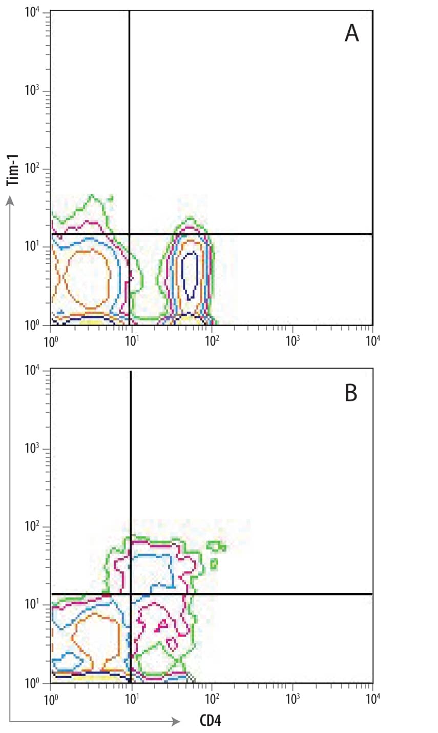
Detection of TIM‑1/KIM‑1/HAVCR in Th2-stimulated Human PBMCs by Flow Cytometry.
(A) Unstimulated and (B) Th2-stimulated human PBMCs were stained with Mouse Anti-Human TIM-1/KIM-1/HAVCR Monoclonal Antibody (Catalog # MAB1750) followed by Allophycocyanin-conjugated Anti-Mouse IgG Secondary Antibody (F0101B) and Human CD4 PerCP-conjugated Monoclonal Antibody (FAB3791C). Quadrant markers were set based on control antibody staining (Catalog # MAB0041).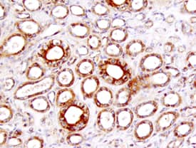
TIM-1/KIM-1/HAVCR in Human Kidney.
TIM-1/KIM-1/HAVCR was detected in immersion fixed paraffin-embedded sections of human kidney using 25 µg/mL Mouse Anti-Human TIM-1/ KIM-1/HAVCR Monoclonal Antibody (Catalog # MAB1750) overnight at 4 °C. Tissue was stained with the Anti-Mouse HRP-DAB Cell & Tissue Staining Kit (brown; CTS002) and counterstained with hematoxylin (blue). View our protocol for Chromogenic IHC Staining of Paraffin-embedded Tissue Sections.Applications for Human TIM-1/KIM-1/HAVCR Antibody
Application
Recommended Usage
CyTOF-ready
Ready to be labeled using established conjugation methods. No BSA or other carrier proteins that could interfere with conjugation.
Flow Cytometry
2.5 µg/106 cells
Sample: stimulated Human CD4+ cells
Sample: stimulated Human CD4+ cells
Immunohistochemistry
8-25 µg/mL
Sample: Immersion fixed paraffin-embedded sections of human kidney
Sample: Immersion fixed paraffin-embedded sections of human kidney
Western Blot
1 µg/mL
Sample: Human CD4+ cells treated with Hamster Anti-Mouse CD3 epsilon Monoclonal Antibody (Catalog # MAB484) and Rat Anti-Mouse CD28 Monoclonal Antibody (Catalog # MAB4831)
Sample: Human CD4+ cells treated with Hamster Anti-Mouse CD3 epsilon Monoclonal Antibody (Catalog # MAB484) and Rat Anti-Mouse CD28 Monoclonal Antibody (Catalog # MAB4831)
Reviewed Applications
Read 4 reviews rated 4.8 using MAB1750 in the following applications:
Formulation, Preparation, and Storage
Purification
Protein A or G purified from hybridoma culture supernatant
Reconstitution
Reconstitute at 0.5 mg/mL in sterile PBS. For liquid material, refer to CoA for concentration.
Formulation
Lyophilized from a 0.2 μm filtered solution in PBS with Trehalose. See Certificate of Analysis for details.
*Small pack size (-SP) is supplied either lyophilized or as a 0.2 µm filtered solution in PBS.
*Small pack size (-SP) is supplied either lyophilized or as a 0.2 µm filtered solution in PBS.
Shipping
Lyophilized product is shipped at ambient temperature. Liquid small pack size (-SP) is shipped with polar packs. Upon receipt, store immediately at the temperature recommended below.
Stability & Storage
Use a manual defrost freezer and avoid repeated freeze-thaw cycles.
- 12 months from date of receipt, -20 to -70 °C as supplied.
- 1 month, 2 to 8 °C under sterile conditions after reconstitution.
- 6 months, -20 to -70 °C under sterile conditions after reconstitution.
Background: TIM-1/KIM-1/HAVCR
TIM-1 (T cell-immunoglobulin-mucin; also KIM-1 and HAVcr-1) is a 100 kDa, type I transmembrane glycoprotein member of the TIM family of immunoglobulin superfamily molecules (1-3). This gene family is involved in the regulation of Th1 and Th2-cell-mediated immunity. Human TIM-1 is synthesized as a 359 amino acid (aa) precursor that contains a 20 aa signal sequence, a 270 aa extracellular domain (ECD), a 21 aa transmembrane segment and a 48 aa cytoplasmic domain (4-6). The ECD contains oneV-type Ig-like domain and a mucin region characterized by multiple PTTTTL motifs. The mucin region undergoes extensive O-linked glycosylation. The TIM-1 gene is highly polymorphic and undergoes alternate splicing (1). For instance, the presence of a six aa sequence (MTTTVP) at position #137 of the mature molecule is associated with protection from atopy in people with a history of hepatitis A (7, 8). There are two cytoplasmic alternate splice forms of
TIM‑1. One is a long (359 aa) kidney form termed TIM-1b, and one is a short (334 aa) liver form termed TIM-1a. Both are identical through the first 323 aa of their precursors. TIM-1b contains a tyrosine phosphorylation motif that is not present in 1a (6). TIM-1 is also known to circulate as a soluble form. Constitutive cleavage by an undefined MMP (possibly ADAM33) releases an 85 - 90 kDa soluble molecule (6). The ECD of human TIM-1 is 50% and 43% aa identical to mouse and canine TIM-1 ECD, respectively. The only two reported ligands for TIM-1 are TIM-4 and the hepatitis A virus (4, 9). However, others are believed to exist, and based on the ligand for TIM-3, one may well be an S-type lectin (10). TIM-1 ligation induces T cell proliferation and promotes cytokine production (1, 10).
References
- Meyers, J.H. et al. (2005) Trends Mol. Med. 11:1471.
- Kuchroo, V.K. et al. (2003) Nat. Rev. Immunol. 3:454.
- Mariat, C. et al. (2005) Phil. Trans. R. Soc. B 360:1681.
- Feigelstock, D. et al. (1998) J. Virol. 72:6621.
- Ichimura, T. et al. (1998) J. Biol. Chem. 273:4135.
- Bailly, V. et al. (2002) J. Biol. Chem. 277:39739.
- Umetsu, D.T. et al. (2005) J. Pediatr. Gastroenterol. Nutr. 40:S43.
- Gao, P-S. et al. (2005) J. Allergy Clin. Immunol. 115:982.
- Zhu, C. et al. (2005) Nat. Immunol. 6:1245.
- Meyers, J.H. et al. (2005) Nat. Immunol. 6:455.
Long Name
T Cell Immunoglobulin Mucin-1
Alternate Names
CD365, HAVCR1, KIM-1, TIM1
Gene Symbol
HAVCR1
UniProt
Additional TIM-1/KIM-1/HAVCR Products
Product Documents for Human TIM-1/KIM-1/HAVCR Antibody
Product Specific Notices for Human TIM-1/KIM-1/HAVCR Antibody
This product is covered by one or more of the following US Patents 7,300,652; 7,041,290; 6,664,385 and other US and foreign patents pending or issued.
For research use only
Loading...
Loading...
Loading...
Loading...
Loading...
Loading...