
Human Siglec-3/CD33 Antibody
R&D Systems, part of Bio-Techne | Catalog # MAB11371

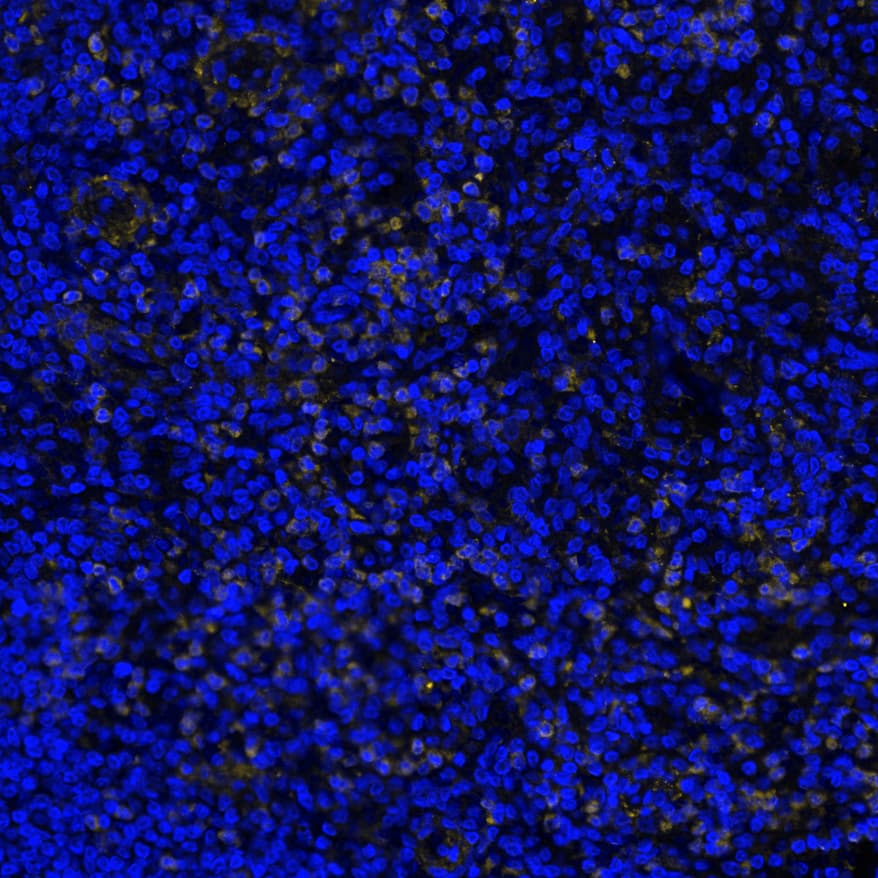
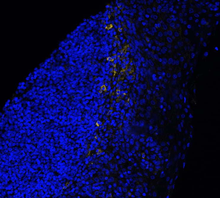
Scientific images may be resized, cropped, or adjusted for brightness or contrast for presentation purposes. These updates are limited to presentation and do not affect the underlying scientific interpretation of the data. Where available, additional source data may be provided upon request.
Conjugate
Catalog #
Key Product Details
Species Reactivity
Validated:
Human
Cited:
Mouse
Applications
Validated:
Immunohistochemistry, Western Blot, Immunocytochemistry, COMET
Cited:
Immunohistochemistry
Label
Unconjugated
Antibody Source
Monoclonal Mouse IgG2B Clone # 996810
Product Specifications
Immunogen
Mouse myeloma cell line, NS0-derived human Siglec-3/CD33
Asp18-His259
Accession # P20138
Asp18-His259
Accession # P20138
Specificity
Detects human Siglec-3/CD33 in direct ELISAs.
Clonality
Monoclonal
Host
Mouse
Isotype
IgG2B
Scientific Data Images for Human Siglec-3/CD33 Antibody
CD33 in Human Tonsil via seqIF™ staining on COMET™
CD33 was detected in immersion fixed paraffin-embedded sections of human tonsil using Mouse Anti-Human CD33, Monoclonal Antibody (Catalog # MAB11371) at 15ug/mL at 37 ° Celsius for 4 minutes. Before incubation with the primary antibody, tissue underwent an all-in-one dewaxing and antigen retrieval preprocessing using PreTreatment Module (PT Module) and Dewax and HIER Buffer H (pH 9; Epredia Catalog # TA-999-DHBH). Tissue was stained using the Alexa Fluor™ 647 Goat anti-Mouse IgG Secondary Antibody at 1:200 at 37 ° Celsius for 2 minutes. (Yellow; Lunaphore Catalog # DR647MS) and counterstained with DAPI (blue; Lunaphore Catalog # DR100). Specific staining was localized to the membrane. Protocol available in COMET™ Panel Builder.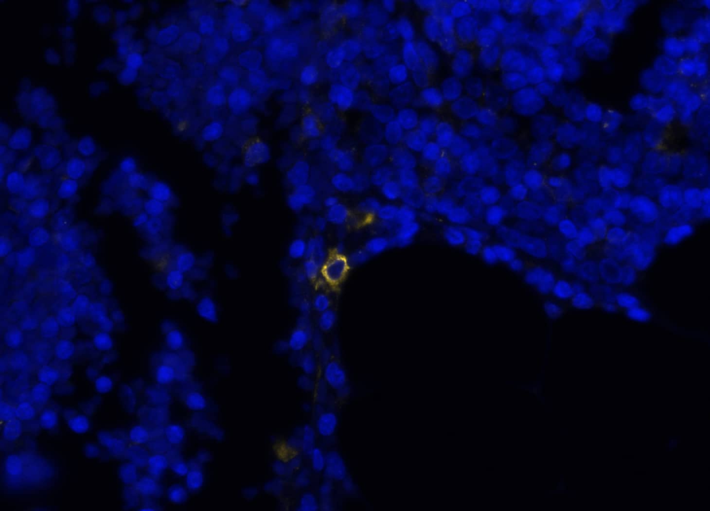
CD33 in Human Thymus via seqIF™ staining on COMET™
CD33 was detected in immersion fixed paraffin-embedded sections of human thymus using Mouse Anti-Human CD33, Monoclonal Antibody (Catalog # MAB11371) at 15ug/mL at 37 ° Celsius for 4 minutes. Before incubation with the primary antibody, tissue underwent an all-in-one dewaxing and antigen retrieval preprocessing using PreTreatment Module (PT Module) and Dewax and HIER Buffer H (pH 9; Epredia Catalog # TA-999-DHBH). Tissue was stained using the Alexa Fluor™ 647 Goat anti-Mouse IgG Secondary Antibody at 1:200 at 37 ° Celsius for 2 minutes. (Yellow; Lunaphore Catalog # DR647MS) and counterstained with DAPI (blue; Lunaphore Catalog # DR100). Specific staining was localized to the membrane. Protocol available in COMET™ Panel Builder.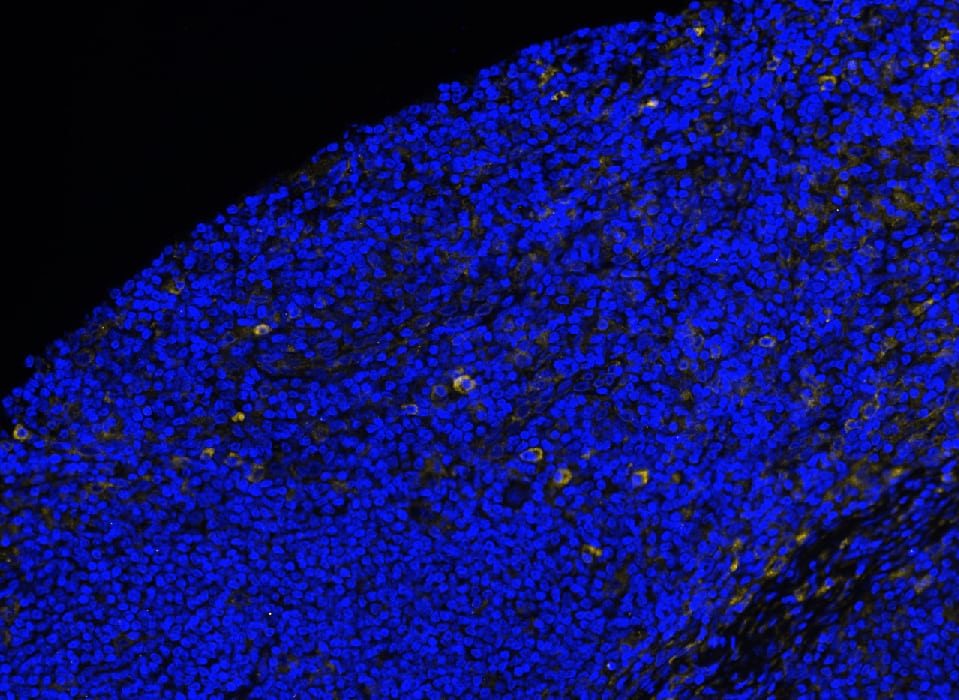
CD33 in Human Hodgkin Lymphoma via seqIF™ staining on COMET™
CD33 was detected in immersion fixed paraffin-embedded sections of human Hodgkin Lymphoma using Mouse Anti-Human CD33, Monoclonal Antibody (Catalog # MAB11371) at 15ug/mL at 37 ° Celsius for 4 minutes. Before incubation with the primary antibody, tissue underwent an all-in-one dewaxing and antigen retrieval preprocessing using PreTreatment Module (PT Module) and Dewax and HIER Buffer H (pH 9; Epredia Catalog # TA-999-DHBH). Tissue was stained using the Alexa Fluor™ 647 Goat anti-Mouse IgG Secondary Antibody at 1:200 at 37 ° Celsius for 2 minutes. (Yellow; Lunaphore Catalog # DR647MS) and counterstained with DAPI (blue; Lunaphore Catalog # DR100). Specific staining was localized to the membrane. Protocol available in COMET™ Panel Builder.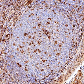

Applications for Human Siglec-3/CD33 Antibody
Application
Recommended Usage
COMET
15 µg/mL
Sample: Immersion fixed paraffin-embedded sections of human tonsil, thymus, Hodgkin Lymphoma and lymph node
Sample: Immersion fixed paraffin-embedded sections of human tonsil, thymus, Hodgkin Lymphoma and lymph node
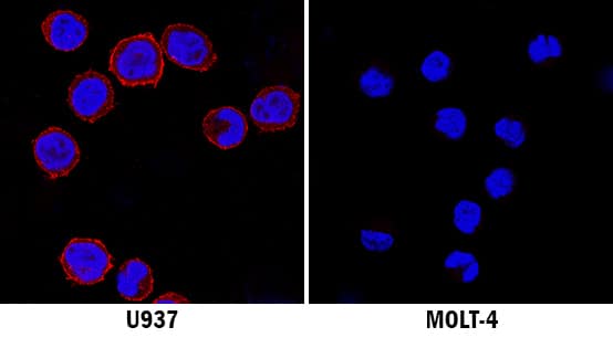
Immunocytochemistry
8-25 µg/mL
Sample: Immersion fixed U937 human histiocytic lymphoma cell line
Sample: Immersion fixed U937 human histiocytic lymphoma cell line
Immunohistochemistry
5-25 µg/mL
Sample: Immersion fixed paraffin-embedded sections of human tonsil
Sample: Immersion fixed paraffin-embedded sections of human tonsil
Western Blot
2 µg/mL
Sample: U937 human histiocytic lymphoma cell line
Sample: U937 human histiocytic lymphoma cell line
Reviewed Applications
Read 4 reviews rated 4.8 using MAB11371 in the following applications:
Formulation, Preparation, and Storage
Purification
Protein A or G purified from hybridoma culture supernatant
Reconstitution
Reconstitute at 0.5 mg/mL in sterile PBS. For liquid material, refer to CoA for concentration.
Formulation
Lyophilized from a 0.2 μm filtered solution in PBS with Trehalose. See Certificate of Analysis for details.
*Small pack size (-SP) is supplied either lyophilized or as a 0.2 µm filtered solution in PBS.
*Small pack size (-SP) is supplied either lyophilized or as a 0.2 µm filtered solution in PBS.
Shipping
Lyophilized product is shipped at ambient temperature. Liquid small pack size (-SP) is shipped with polar packs. Upon receipt, store immediately at the temperature recommended below.
Stability & Storage
Use a manual defrost freezer and avoid repeated freeze-thaw cycles.
- 12 months from date of receipt, -20 to -70 °C as supplied.
- 1 month, 2 to 8 °C under sterile conditions after reconstitution.
- 6 months, -20 to -70 °C under sterile conditions after reconstitution.
Background: Siglec-3/CD33
Human Siglec-3 is alternatively known as myeloid cell surface antigen CD33 and GP67. Human Siglec-3 cDNA encodes a 364 amino acid (aa) polypeptide with a hydrophobic signal peptide, an N-terminal Ig-like V-type domain, one Ig-like C2-type domains, a transmembrane region and a cytoplasmic tail (1, 4). Siglec-3 expression is restricted to cells of myelomonocytic lineage (2). It binds sialic acid preferring alpha2,3- linkage over alpha2,6- linkage (5). Studies indicated that Siglec-3 recruits SHP-1 and SHP-2 to its ITIMs (6, 7). When co-crosslinking with Fc gammaR1, Siglec-3 inhibits tyrosine phosphorylation and calcium mobilization, suggesting Siglec-3 can mediate inhibitory signals (7).
References
- Crocker, P.R. and A. Varki (2001) Trends Immunol. 22:337.
- Crocker, P.R. and A. Varki (2001) Immunology 103:137.
- Angata, T. et al. (2002) J. Biol. Chem. 277:24466.
- Simmons, D. and B. Seed (1988) J. Immunol. 141:2797.
- Freeman, S.D. et al. (1995) Blood 85:2002.
- Taylor, V.C. et al. (1999) J. Biol. Chem. 274:11505.
- Ulyanova, T. et al. (1999) Eur. J. Immunol. 29:3440.
Long Name
Sialic Acid Binding Ig-like Lectin 3
Alternate Names
CD33, gp67, Siglec3
Gene Symbol
CD33
UniProt
Additional Siglec-3/CD33 Products
Product Documents for Human Siglec-3/CD33 Antibody
Product Specific Notices for Human Siglec-3/CD33 Antibody
For research use only
Loading...
Loading...
Loading...
Loading...
Loading...