
Human/Mouse/Rat AMPK beta1 Antibody
R&D Systems, part of Bio-Techne | Catalog # AF2854


Conjugate
Catalog #
Key Product Details
Species Reactivity
Human, Mouse, Rat
Applications
Immunohistochemistry, Western Blot
Label
Unconjugated
Antibody Source
Polyclonal Goat IgG
Product Specifications
Immunogen
E. coli-derived recombinant human AMPK beta1
Met1-Ile270
Accession # Q9Y478
Met1-Ile270
Accession # Q9Y478
Specificity
Detects human, mouse, and rat AMPK beta1 in Western blots. In Western blots, no cross-reactivity with recombinant human AMPK beta2 is observed.
Clonality
Polyclonal
Host
Goat
Isotype
IgG
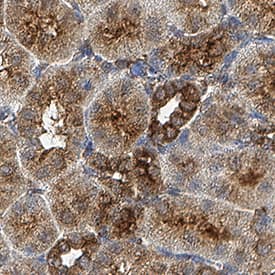
Scientific Data Images for Human/Mouse/Rat AMPK beta1 Antibody
Detection of Human/Mouse/Rat AMPK beta1 by Western Blot.
Western blot shows lysates of HepG2 human hepatocellular carcinoma cell line and C6 rat glioma cell line. PVDF membrane was probed with 1 µg/mL Goat Anti-Human/Mouse/Rat AMPK beta1 Antigen Affinity-purified Polyclonal Antibody (Catalog # AF2854) followed by HRP-conjugated Anti-Goat IgG Secondary Antibody (HAF109). For additional reference, recombinant human AMPK beta1 and AMPK beta2 (2 ng/lane) were included. A specific band for AMPK beta 1 was detected at approximately 38 kDa (as indicated). This experiment was conducted under reducing conditions and using Immunoblot Buffer Group 2.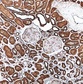
Detection of AMPK beta1 in Human Kidney.
AMPK beta1 was detected in immersion fixed paraffin-embedded sections of Human Kidney using Goat Anti-Human/Mouse/Rat AMPK beta1 Antigen Affinity-purified Polyclonal Antibody (Catalog # AF2854) at 3 µg/mL for 1 hour at room temperature followed by incubation with the Anti-Goat IgG VisUCyte™ HRP Polymer Antibody (Catalog # VC004). Before incubation with the primary antibody, tissue was subjected to heat-induced epitope retrieval using VisUCyte Antigen Retrieval Reagent-Basic (Catalog # VCTS021). Tissue was stained using DAB (brown) and counterstained with hematoxylin (blue). Specific staining was localized to cytoplasm in convoluted tubules. View our protocol for IHC Staining with VisUCyte HRP Polymer Detection Reagents.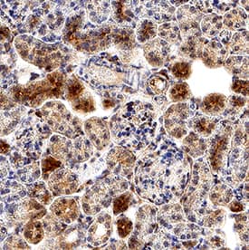
Detection of AMPK beta1 in Mouse Kidney.
AMPK beta1 was detected in immersion fixed paraffin-embedded sections of Mouse Kidney using Goat Anti-Human/Mouse/Rat AMPK beta1 Antigen Affinity-purified Polyclonal Antibody (Catalog # AF2854) at 3 µg/mL for 1 hour at room temperature followed by incubation with the Anti-Goat IgG VisUCyte™ HRP Polymer Antibody (Catalog # VC004). Before incubation with the primary antibody, tissue was subjected to heat-induced epitope retrieval using VisUCyte Antigen Retrieval Reagent-Basic (Catalog # VCTS021). Tissue was stained using DAB (brown) and counterstained with hematoxylin (blue). Specific staining was localized to cytoplasm in convoluted tubules. View our protocol for IHC Staining with VisUCyte HRP Polymer Detection Reagents.Applications for Human/Mouse/Rat AMPK beta1 Antibody
Application
Recommended Usage
Immunohistochemistry
3-15 µg/mL
Sample: Immersion fixed paraffin-embedded sections of Human Kidney, Mouse Kidney and Rat Kidney
Sample: Immersion fixed paraffin-embedded sections of Human Kidney, Mouse Kidney and Rat Kidney
Western Blot
1 µg/mL
Sample: HepG2 human hepatocellular carcinoma cell line and C6 rat glioma cell line
Sample: HepG2 human hepatocellular carcinoma cell line and C6 rat glioma cell line
Reviewed Applications
Read 1 review rated 4 using AF2854 in the following applications:
Formulation, Preparation, and Storage
Purification
Antigen Affinity-purified
Reconstitution
Reconstitute at 0.2 mg/mL in sterile PBS. For liquid material, refer to CoA for concentration.
Formulation
Lyophilized from a 0.2 μm filtered solution in PBS with Trehalose. *Small pack size (SP) is supplied either lyophilized or as a 0.2 µm filtered solution in PBS.
Shipping
Lyophilized product is shipped at ambient temperature. Liquid small pack size (-SP) is shipped with polar packs. Upon receipt, store immediately at the temperature recommended below.
Stability & Storage
Use a manual defrost freezer and avoid repeated freeze-thaw cycles.
- 12 months from date of receipt, -20 to -70 °C as supplied.
- 1 month, 2 to 8 °C under sterile conditions after reconstitution.
- 6 months, -20 to -70 °C under sterile conditions after reconstitution.
Background: AMPK beta 1
AMP-activated protein kinase (AMPK) is a heterotrimeric complex consisting of a catalytic alpha subunit and regulatory beta and gamma subunits. Each subunit exists as alternate isoforms ( alpha1, alpha2, beta1, beta2, gamma1, gamma2, gamma3), with all 12 combinations able to form complexes. The beta1 subunit is expressed at higher levels than the beta2 subunit in liver, while beta2 is more abundant than beta1 in skeletal muscle. AMPK’s role in metabolic regulation has implicated this serine/threonine kinase complex as a therapeutic target in heart disease, obesity, and diabetes.
Long Name
AMP-activated Protein Kinase beta 1
Alternate Names
PRKAB1
Gene Symbol
PRKAB1
UniProt
Additional AMPK beta 1 Products
Product Documents for Human/Mouse/Rat AMPK beta1 Antibody
Product Specific Notices for Human/Mouse/Rat AMPK beta1 Antibody
For research use only
Loading...
Loading...
Loading...
Loading...
Loading...