
Human LAP (TGF-beta 1) Antibody
R&D Systems, part of Bio-Techne | Catalog # AF-246-NA

Scientific images may be resized, cropped, or adjusted for brightness or contrast for presentation purposes. These updates are limited to presentation and do not affect the underlying scientific interpretation of the data. Where available, additional source data may be provided upon request.
Conjugate
Catalog #
Key Product Details
Species Reactivity
Validated:
Human
Cited:
Human, Mouse, Rat, Porcine, Mink, Rabbit, Transgenic Mouse
Applications
Validated:
Western Blot, Neutralization, Immunocytochemistry, Simple Western
Cited:
Immunohistochemistry, Immunohistochemistry-Paraffin, Immunohistochemistry-Frozen, Western Blot, Neutralization, Immunocytochemistry, Bioassay, Dot Blot
Label
Unconjugated
Antibody Source
Polyclonal Goat IgG
Product Specifications
Immunogen
S. frugiperda insect ovarian cell line Sf 21-derived and Chinese hamster ovary cell line CHO-derived recombinant human LAP TGF‑ beta1
Leu30-Ser390
Accession # P01137
Leu30-Ser390
Accession # P01137
Specificity
Detects human LAP TGF‑ beta1 in direct ELISAs and Western blots.
Clonality
Polyclonal
Host
Goat
Isotype
IgG
Endotoxin Level
<0.10 EU per 1 μg of the antibody by the LAL method.
Scientific Data Images for Human LAP (TGF-beta 1) Antibody
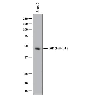
Detection of Human LAP (TGF-beta 1) by Western Blot.
Western blot shows lysates of Saos-2 human osteosarcoma cell line. PVDF membrane was probed with 2 µg/mL of Goat Anti-Human LAP (TGF-beta 1) Antigen Affinity-purified Polyclonal Antibody (Catalog # AF-246-NA) followed by HRP-conjugated Anti-Goat IgG Secondary Antibody (Catalog # HAF017). A specific band was detected for LAP (TGF-beta 1) at approximately 48 kDa (as indicated). This experiment was conducted under reducing conditions and using Immunoblot Buffer Group 1.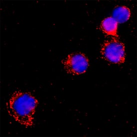
LAP (TGF-beta 1) in Human PBMCs.
LAP (TGF-beta 1) was detected in immersion fixed Human peripheral blood mononuclear cells using Goat Anti-Human LAP (TGF-beta 1) Antigen Affinity-purified Polyclonal Antibody (Catalog # AF-246-NA) at 15 µg/mL for 3 hours at room temperature. Cells were stained using the NorthernLights™ 557-conjugated Anti-Goat IgG Secondary Antibody (red; NL001) and counterstained with DAPI (blue). Specific staining was localized to cytoplasm. Staining was performed using our protocol for Fluorescent ICC Staining of Non-adherent Cells.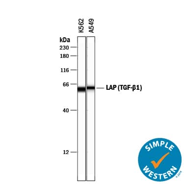
Detection of Human LAP (TGF-beta 1) by Simple WesternTM.
Simple Western lane view shows lysates of K562 human chronic myelogenous leukemia cell line and A549 human lung carcinoma cell line, loaded at 0.2 mg/mL. A specific band was detected for LAP (TGF-beta 1) at approximately 61 kDa (as indicated) using 20 µg/mL of Goat Anti-Human LAP (TGF-beta 1) Antigen Affinity-purified Polyclonal Antibody (Catalog # AF-246-NA) followed by 1:50 dilution of HRP-conjugated Anti-Goat IgG Secondary Antibody (Catalog # HAF109). This experiment was conducted under reducing conditions and using the 12-230 kDa separation system.Applications for Human LAP (TGF-beta 1) Antibody
Application
Recommended Usage
Immunocytochemistry
5-15 µg/mL
Sample: Immersion fixed Human peripheral blood mononuclear cells
Sample: Immersion fixed Human peripheral blood mononuclear cells
Simple Western
20 µg/mL
Sample: K562 human chronic myelogenous leukemia cell line and A549 human lung carcinoma cell line
Sample: K562 human chronic myelogenous leukemia cell line and A549 human lung carcinoma cell line
Western Blot
2 µg/mL
Sample: Saos‑2 human osteosarcoma cell line
Sample: Saos‑2 human osteosarcoma cell line
Neutralization
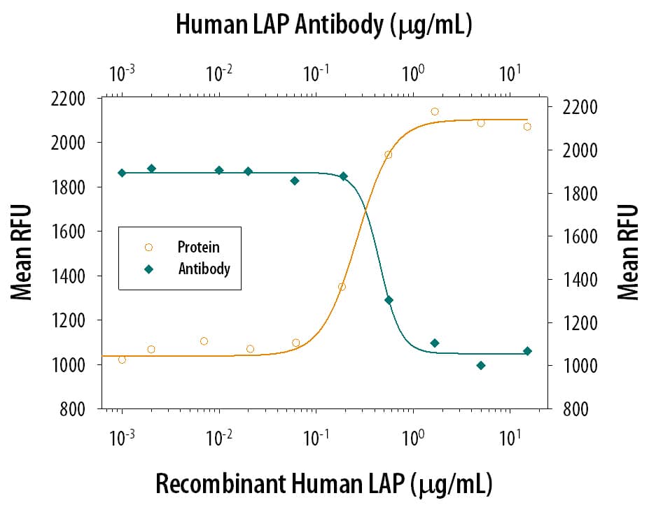
Measured by its ability to neutralize LAP TGF‑ beta1 inhibition of TGF‑ beta1 growth inhibition in the HT‑2 mouse T cell line. Tsang, M. et al. (1995) Cytokine 7:389. The Neutralization Dose (ND50) is typically 0.4-2 µg/mL in the presence of 500 ng/mL Recombinant Human LAP TGF‑ beta1 and 1 ng/mL TGF‑ beta1.
Reviewed Applications
Read 4 reviews rated 4 using AF-246-NA in the following applications:
Formulation, Preparation, and Storage
Purification
Antigen Affinity-purified
Reconstitution
Reconstitute at 0.2 mg/mL in sterile PBS. For liquid material, refer to CoA for concentration.
Formulation
Lyophilized from a 0.2 μm filtered solution in PBS with Trehalose. *Small pack size (SP) is supplied either lyophilized or as a 0.2 µm filtered solution in PBS.
Shipping
Lyophilized product is shipped at ambient temperature. Liquid small pack size (-SP) is shipped with polar packs. Upon receipt, store immediately at the temperature recommended below.
Stability & Storage
Use a manual defrost freezer and avoid repeated freeze-thaw cycles.
- 12 months from date of receipt, -20 to -70 °C as supplied.
- 1 month, 2 to 8 °C under sterile conditions after reconstitution.
- 6 months, -20 to -70 °C under sterile conditions after reconstitution.
Background: LAP (TGF-beta 1)
TGF-beta 1 (transforming growth factor beta 1) and the closely related TGF-beta 2 and -beta 3 are members of the large TGF-beta superfamily. TGF‑ beta proteins are highly pleiotropic cytokines that regulate processes such as immune function, proliferation and epithelial-mesenchymal transition (1‑3). Human TGF-beta 1 cDNA encodes a 390 amino acid (aa) precursor that contains a 29 aa signal peptide and a 361 aa proprotein (4). A furin-like convertase processes the proprotein within the trans-Golgi to generate an N‑terminal 249 aa (aa 30-278) latency-associated peptide (LAP) and a C-terminal 112 aa mature TGF-beta 1 (aa 279-390) (4‑6). Disulfide-linked homodimers of LAP and TGF-beta 1 remain non‑covalently associated after secretion, forming the small latent TGF-beta 1 complex (4‑8). Purified LAP is also capable of associating with active TGF-beta with high affinity, and can neutralize TGF-beta activity (9). Covalent linkage of LAP to one of three latent TGF-beta binding proteins (LTBPs) creates a large latent complex that may interact with the extracellular matrix (5-7). TGF-beta activation from latency is controlled both spatially and temporally, by multiple pathways that include actions of proteases such as plasmin and MMP9, and/or by thrombospondin 1 or selected integrins (5, 8). The LAP portion of human TGF-beta 1 shares 91%, 92%, 85%, 86% and 88% aa identity with porcine, canine, mouse, rat and equine TGF-beta 1 LAP, respectively, while mature human TGF-beta 1 portion shares 100% aa identity with porcine, canine and bovine TGF-beta 1, and 99% aa identity with mouse, rat and equine TGF-beta 1. Although different isoforms of TGF-beta are naturally associated with their own distinct LAPs, the TGF-beta 1 LAP is capable of complexing with, and inactivating, all other human TGF-beta isoforms and those of most other species (9). Mutations within the LAP are associated with Camurati-Engelmann disease, a rare sclerosing bone dysplasia characterized by inappropriate presence of active TGF-beta 1 (10).
References
- Dunker, N. & K. Krieglstein (2000) Eur. J. Biochem. 267:6982.
- Wahl, S.M. (2006) Immunol. Rev. 213:213.
- Chang, H. et al. (2002) Endocr. Rev. 23:787.
- Derynck, R. et al. (1985) Nature 316:701.
- Dabovic, B. and D.B. Rifkin (2008) “TGF-beta Bioavailability” in The TGF-beta Family. Derynck, R. and K. Miyazono (eds): Cold Spring Harbor Laboratory Press, p. 179.
- Brunner, A.M. et al. (1989) J. Biol. Chem. 264:13660.
- Miyazono, K. et al. (1991) EMBO J. 10:1091.
- Oklu, R. and R. Hesketh (2000) Biochem. J. 352:601.
- Miller, D.M. et al. (1992) Mol. Endocrinol. 6:694.
- Janssens, K. et al. (2003) J. Biol. Chem. 278:7718.
Long Name
Latency-associated Peptide
Alternate Names
LAP (TGFbeta 1)
Entrez Gene IDs
7040 (Human)
Gene Symbol
TGFB1
UniProt
Additional LAP (TGF-beta 1) Products
Product Documents for Human LAP (TGF-beta 1) Antibody
Product Specific Notices for Human LAP (TGF-beta 1) Antibody
For research use only
Loading...
Loading...
Loading...
Loading...
Loading...