
Human IL-4R alpha Antibody
R&D Systems, part of Bio-Techne | Catalog # MAB230



Scientific images may be resized, cropped, or adjusted for brightness or contrast for presentation purposes. These updates are limited to presentation and do not affect the underlying scientific interpretation of the data. Where available, additional source data may be provided upon request.
Conjugate
Catalog #
Key Product Details
Species Reactivity
Validated:
Human
Cited:
Human, Mouse, Rat
Applications
Validated:
Immunohistochemistry, Western Blot, Neutralization, Flow Cytometry, Immunocytochemistry, CyTOF-ready
Cited:
Immunohistochemistry, Immunohistochemistry-Paraffin, Immunohistochemistry-Frozen, Western Blot, Neutralization, Flow Cytometry, Immunocytochemistry, Binding Assay, Bioassay, Blocking, In vivo assay
Label
Unconjugated
Antibody Source
Monoclonal Mouse IgG2A Clone # 25463
Product Specifications
Immunogen
S. frugiperda insect ovarian cell line Sf 21-derived recombinant human IL-4 R
Gly24-His232
Accession # P24394
Gly24-His232
Accession # P24394
Specificity
Detects human IL‑4 R alpha in direct ELISAs and Western blots. In direct ELISAs, no cross-reactivity with recombinant human (rh) IL-9 R, recombinant mouse IL-4 R, rhIL-5 R alpha, rhIL-5 R beta, rhIL-13 R alpha1, or rhIL-13 R alpha2 is observed.
Clonality
Monoclonal
Host
Mouse
Isotype
IgG2A
Endotoxin Level
<0.10 EU per 1 μg of the antibody by the LAL method.
Scientific Data Images for Human IL-4R alpha Antibody
Detection of IL‑4 R alpha in Daudi Human Cell Line by Flow Cytometry.
Daudi human Burkitt's lymphoma cell line was stained with Mouse Anti-Human IL-4 Ra Monoclonal Antibody (Catalog # MAB230, filled histogram) or isotype control antibody (Catalog # MAB003, open histogram), followed by Phycoerythrin-conjugated Anti-Mouse IgG Secondary Antibody (Catalog # F0102B).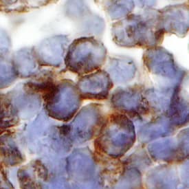
IL‑4 R alpha in Human Lymph Node.
IL-4 Ra was detected in immersion fixed paraffin-embedded sections of human lymph node using Mouse Anti-Human IL-4 Ra Monoclonal Antibody (Catalog # MAB230) at 25 µg/mL overnight at 4 °C. Tissue was stained using the Anti-Mouse HRP-DAB Cell & Tissue Staining Kit (brown; Catalog # CTS002) and counterstained with hematoxylin (blue). Specific staining was localized to plasma cells. View our protocol for Chromogenic IHC Staining of Paraffin-embedded Tissue Sections.
IL‑4 R alpha in HDLM‑2 Human Cell Line.
IL-4 Ra was detected in immersion fixed HDLM-2 human Hodgkin's lymphoma cell line using Mouse Anti-Human IL-4 Ra Monoclonal Antibody (Catalog # MAB230) at 8 µg/mL for 3 hours at room temperature. Cells were stained using the NorthernLights™ 557-conjugated Anti-Mouse IgG Secondary Antibody (red; Catalog # NL007) and counterstained with DAPI (blue). Specific staining was localized to cytoplasm. View our protocol for Fluorescent ICC Staining of Non-adherent Cells.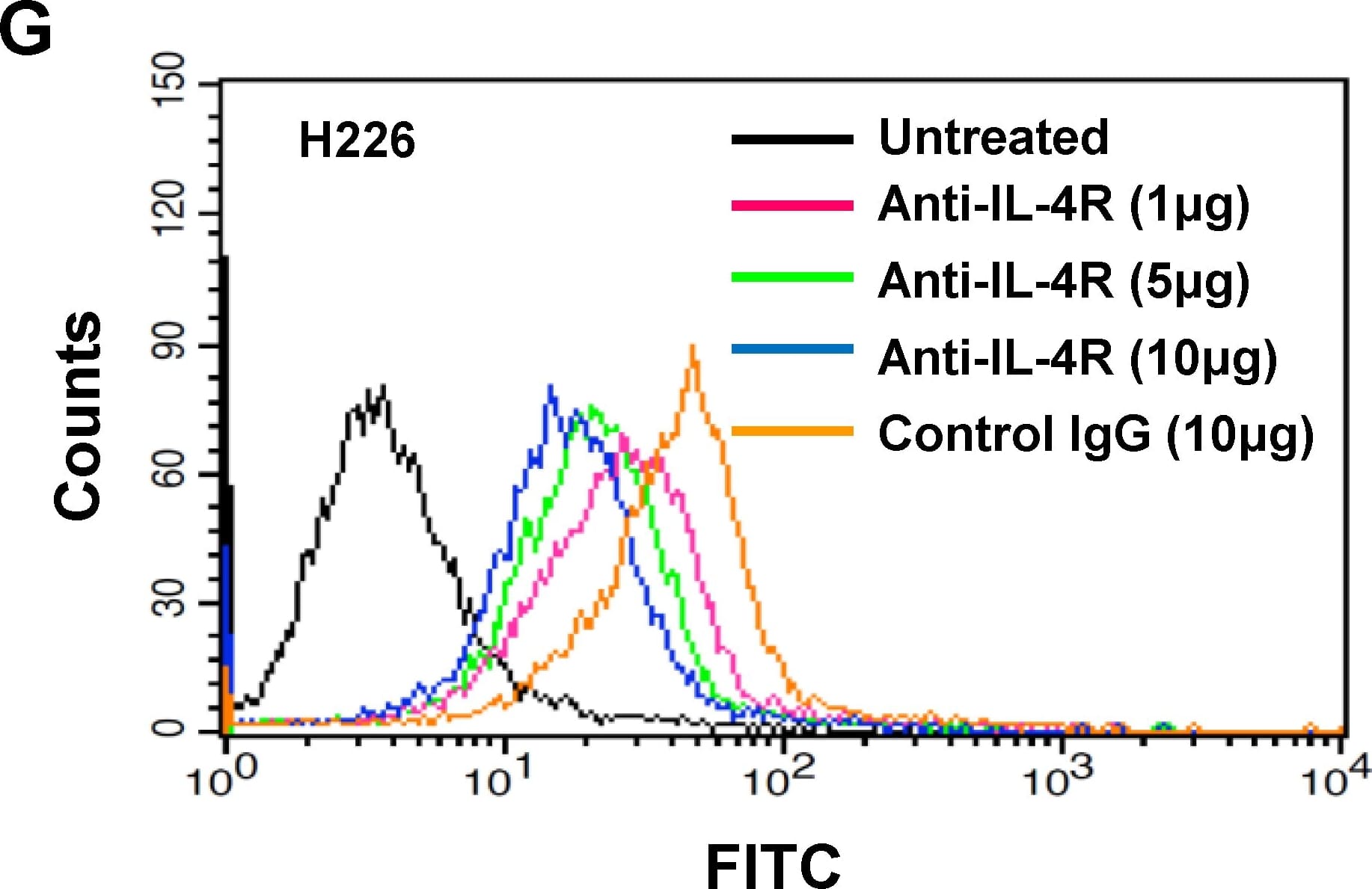
Applications for Human IL-4R alpha Antibody
Application
Recommended Usage
CyTOF-ready
Ready to be labeled using established conjugation methods. No BSA or other carrier proteins that could interfere with conjugation.
Flow Cytometry
0.25 µg/106 cells
Sample: Daudi human Burkitt's lymphoma cell line
Sample: Daudi human Burkitt's lymphoma cell line
Immunocytochemistry
5-15 µg/mL
Sample: Immersion fixed HDLM-2 human Hodgkin's lymphoma cell line
Sample: Immersion fixed HDLM-2 human Hodgkin's lymphoma cell line
Immunohistochemistry
8-25 µg/mL
Sample: Immersion fixed paraffin-embedded sections of human lymph node
Sample: Immersion fixed paraffin-embedded sections of human lymph node
Western Blot
1 µg/mL
Sample: Recombinant Human IL‑4 R alpha (Catalog # 230-4R) under non-reducing conditions only
Sample: Recombinant Human IL‑4 R alpha (Catalog # 230-4R) under non-reducing conditions only
Neutralization
Measured by its ability to neutralize IL-4-induced proliferation in the TF-1 human erythroleukemic cell line. Kitamura, T. et al. (1989) J. Cell Physiol. 140:323. The Neutralization Dose (ND50) is typically 3-6 ng/mL in the presence of 0.2 ng/mL Recombinant Human IL-4.
Reviewed Applications
Read 4 reviews rated 4.5 using MAB230 in the following applications:
Formulation, Preparation, and Storage
Purification
Protein A or G purified from ascites
Reconstitution
Reconstitute at 0.5 mg/mL in sterile PBS. For liquid material, refer to CoA for concentration.
Formulation
Lyophilized from a 0.2 μm filtered solution in PBS with Trehalose. *Small pack size (SP) is supplied either lyophilized or as a 0.2 µm filtered solution in PBS.
Shipping
Lyophilized product is shipped at ambient temperature. Liquid small pack size (-SP) is shipped with polar packs. Upon receipt, store immediately at the temperature recommended below.
Stability & Storage
Use a manual defrost freezer and avoid repeated freeze-thaw cycles.
- 12 months from date of receipt, -20 to -70 °C as supplied.
- 1 month, 2 to 8 °C under sterile conditions after reconstitution.
- 6 months, -20 to -70 °C under sterile conditions after reconstitution.
Background: IL-4R alpha
Interleukin 4 receptor (IL-4 R) is the transmembrane ligand binding subunit of the heterodimeric IL-4 receptor complex. In the type I receptor complex, IL-4 R associates with the common gamma chain, whereas in the type II receptor complex, it associates with IL-13 R alpha1. IL-4 R is expressed by B cells, T cells, monocytes and macrophages.
Long Name
Interleukin 4 Receptor alpha
Alternate Names
CD124, IL-4 R alpha, IL-4Ra, IL4R, IL4R alpha
Gene Symbol
IL4R
UniProt
Additional IL-4R alpha Products
Product Documents for Human IL-4R alpha Antibody
Product Specific Notices for Human IL-4R alpha Antibody
For research use only
Loading...
Loading...
Loading...
Loading...
Loading...
Loading...