
Human IL-12 R beta2 Antibody
R&D Systems, part of Bio-Techne | Catalog # MAB19591

Scientific images may be resized, cropped, or adjusted for brightness or contrast for presentation purposes. These updates are limited to presentation and do not affect the underlying scientific interpretation of the data. Where available, additional source data may be provided upon request.
Conjugate
Catalog #
Key Product Details
Species Reactivity
Human
Applications
Western Blot
Label
Unconjugated
Antibody Source
Monoclonal Mouse IgG2A Clone # 622509
Product Specifications
Immunogen
Mouse myeloma cell line NS0-derived recombinant human IL-12 R beta2
Lys24-Cys622
Accession # Q99665
Lys24-Cys622
Accession # Q99665
Specificity
Detects human IL-12 R beta2 in direct ELISAs and Western blots. In direct ELISAs, no cross-reactivity with recombinant human IL‑12 R beta1 or recombinant mouse IL-12 R beta1 is observed.
Clonality
Monoclonal
Host
Mouse
Isotype
IgG2A
Scientific Data Images for Human IL-12 R beta2 Antibody
Detection of Human IL‑12 R beta2 by Western Blot.
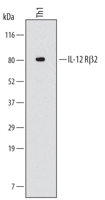
Western blot shows lysates of human CD4+in vitrodifferentiated Th1 cells. PVDF Membrane was probed with 2 µg/mL of Mouse Anti-Human IL-12 R beta2 Monoclonal Antibody (Catalog # MAB19591) followed by HRP-conjugated Anti-Mouse IgG Secondary Antibody (Catalog # HAF007). A specific band was detected for IL-12 R beta2 at approximately 80 kDa (as indicated). This experiment was conducted under reducing conditions and using Immunoblot Buffer Group 1.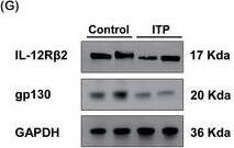
Detection of Human IL-12 R beta2 by Western Blot
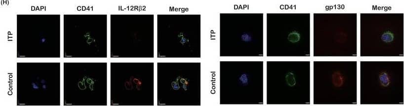
Reduced IL‐35 in patients with ITP. A) The level of plasma IL‐35 by ELISA (n = 12 in the control group, n = 26 in the ITP group). B) The concentration of IL‐35 in the bone marrow of ITP patients and healthy controls by ELISA (n = 22 in the control group, n = 10 in the ITP group). C) Expression of IL‐35 receptor subunit IL‐12R beta2 in CD41/CD61+ cells by flow cytometry (n = 10). D) The mean fluorescence intensity of IL‐12R beta2 on day 10 by flow cytometry (n = 10). E) Expression of IL‐35 receptor subunit gp130 in CD41/CD61+ cells by flow cytometry (n = 10). F) The mean fluorescence intensity of gp130 on day 10 by flow cytometry (n = 10). G) The protein levels of IL‐35 receptor subunits IL‐12R beta2 and gp130 in CD41/CD61+ cells (n = 6). H) CD41 (green), IL‐12R beta2/ gp130 (red), and DAPI (blue) staining of CD41/CD61+ cells (600×; bar: 20 µm). Data information: Data are presented as mean ± SEM. *p < 0.05, **p < 0.01, ***p < 0.001; ns, no significant difference, using Student‘s t‐test (A, B, D, F) or ANOVA (C, E). Image collected and cropped by CiteAb from the following open publication (https://pubmed.ncbi.nlm.nih.gov/38225757), licensed under a CC-BY license. Not internally tested by R&D Systems.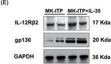
Detection of Human IL-12 R beta2 by Western Blot
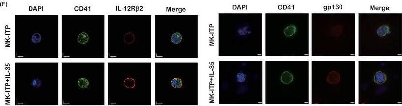
IL‐35 stimulates megakaryopoiesis in vitro. A) Expression of IL‐35 receptor subunit IL‐12R beta2 in CD41/CD61+ cells from ITP patients by flow cytometry (n = 10). B) The mean fluorescence intensity of IL‐12R beta2 on day 10 by flow cytometry (n = 10). C) Expression of IL‐35 receptor subunit gp130 in CD41/CD61+ cells from ITP patients by flow cytometry (n = 10). D) The mean fluorescence intensity of gp130 on day 10 by flow cytometry (n = 10). E) The protein levels of IL‐35 receptor subunits IL‐12R beta2 and gp130 in CD41/CD61+ cells (n = 6). F) CD41 (green), IL‐12R beta2/ gp130 (red), and DAPI (blue) staining of CD41/CD61+ cells (600×; bar: 20 µm). G) (Left) Representative images of megakaryocyte colonies (100×; bar: 100 µm). (Right) The number of megakaryocyte colonies (n = 3). Data information: Data are presented as mean ± SEM. *p < 0.05, **p < 0.01, ***p < 0.001; ns, no significant difference, using ANOVA (A, C, E, F) or Student‘s t‐test (B, D). Image collected and cropped by CiteAb from the following open publication (https://pubmed.ncbi.nlm.nih.gov/38225757), licensed under a CC-BY license. Not internally tested by R&D Systems.Applications for Human IL-12 R beta2 Antibody
Application
Recommended Usage
Western Blot
2 µg/mL
Sample: Human Th1 cells
Sample: Human Th1 cells
Formulation, Preparation, and Storage
Purification
Protein A or G purified from hybridoma culture supernatant
Reconstitution
Sterile PBS to a final concentration of 0.5 mg/mL. For liquid material, refer to CoA for concentration.
Formulation
Lyophilized from a 0.2 μm filtered solution in PBS with Trehalose. *Small pack size (SP) is supplied either lyophilized or as a 0.2 µm filtered solution in PBS.
Shipping
Lyophilized product is shipped at ambient temperature. Liquid small pack size (-SP) is shipped with polar packs. Upon receipt, store immediately at the temperature recommended below.
Stability & Storage
Use a manual defrost freezer and avoid repeated freeze-thaw cycles.
- 12 months from date of receipt, -20 to -70 °C as supplied.
- 1 month, 2 to 8 °C under sterile conditions after reconstitution.
- 6 months, -20 to -70 °C under sterile conditions after reconstitution.
Background: IL-12 R beta 2
IL-12 R beta2 is a type I cytokine receptor family protein that associates with IL 12 R beta1 to form a functional high affinity receptor complex for IL-12. The extracellular region of IL 12 R beta2 contains five fibronectin type 3 domains. The cytoplasmic region is alternatively spliced, giving rise to two isoforms. IL-12 R beta2 is up‑regulated in Th1 cells during inflammatory responses. The extracellular regions of human and mouse IL‑12 R beta2 share 69% amino acid sequence identity.
Long Name
Interleukin 12 Receptor beta 2
Alternate Names
IL-12Rb2, IL12R beta 2, IL12RB2
Gene Symbol
IL12RB2
UniProt
Additional IL-12 R beta 2 Products
Product Documents for Human IL-12 R beta2 Antibody
Product Specific Notices for Human IL-12 R beta2 Antibody
For research use only
Loading...
Loading...
Loading...
Loading...
Loading...