
Human Galectin-3BP/MAC-2BP Antibody
R&D Systems, part of Bio-Techne | Catalog # AF2226


Scientific images may be resized, cropped, or adjusted for brightness or contrast for presentation purposes. These updates are limited to presentation and do not affect the underlying scientific interpretation of the data. Where available, additional source data may be provided upon request.
Conjugate
Catalog #
Key Product Details
Species Reactivity
Validated:
Human
Cited:
Human, Mouse, Primate - Macaca mulatta (Rhesus Macaque)
Applications
Validated:
Western Blot, Immunocytochemistry, Simple Western
Cited:
Immunohistochemistry, Western Blot, Immunocytochemistry
Label
Unconjugated
Antibody Source
Polyclonal Goat IgG
Product Specifications
Immunogen
Mouse myeloma cell line NS0-derived recombinant human Galectin‑3BP/MAC‑2BP
Val19-Asp585
Accession # Q08380
Val19-Asp585
Accession # Q08380
Specificity
Detects human Galectin‑3BP/MAC‑2BP in direct ELISAs and Western blots. In direct ELISAs, less than 20% cross-reactivity with recombinant mouse Galectin‑3BP/MAC‑2BP is observed.
Clonality
Polyclonal
Host
Goat
Isotype
IgG
Scientific Data Images for Human Galectin-3BP/MAC-2BP Antibody
Detection of Human Galectin‑3BP/MAC‑2BP by Western Blot.
Western blot shows lysates of HeLa human cervical epithelial carcinoma cell line and HepG2 human hepatocellular carcinoma cell line. PVDF membrane was probed with 1.0 µg/mL of Goat Anti-Human Galectin-3BP/MAC-2BP Antigen Affinity-purified Polyclonal Antibody (Catalog # AF2226) followed by HRP-conjugated Anti-Goat IgG Secondary Antibody (HAF019). A specific band was detected for Galectin-3BP/MAC-2BP at approximately 75 kDa (as indicated). This experiment was conducted under reducing conditions and using Immunoblot Buffer Group 1.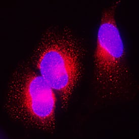
Galectin‑3BP/MAC‑2BP in HeLa Human Cell Line.
Galectin-3BP/MAC-2BP was detected in immersion fixed HeLa human cervical epithelial carcinoma cell line using Goat Anti-Human Galectin-3BP/MAC-2BP Antigen Affinity-purified Polyclonal Antibody (Catalog # AF2226) at 1.7 µg/mL for 3 hours at room temperature. Cells were stained using the NorthernLights™ 557-conjugated Anti-Goat IgG Secondary Antibody (red;NL001) and counterstained with DAPI (blue). Specific staining was localized to cytoplasm. View our protocol for Fluorescent ICC Staining of Cells on Coverslips.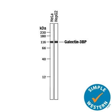
Detection of Human Galectin‑3BP/MAC‑2BP by Simple WesternTM.
Simple Western lane view shows lysates of HeLa human cervical epithelial carcinoma cell line and HepG2 human hepatocellular carcinoma cell line, loaded at 0.2 mg/mL. A specific band was detected for Galectin-3BP/MAC-2BP at approximately 115 kDa (as indicated) using 10 µg/mL of Goat Anti-Human Galectin-3BP/MAC-2BP Antigen Affinity-purified Polyclonal Antibody (Catalog # AF2226) followed by 1:50 dilution of HRP-conjugated Anti-Goat IgG Secondary Antibody (HAF109). This experiment was conducted under reducing conditions and using the 12-230 kDa separation system.Applications for Human Galectin-3BP/MAC-2BP Antibody
Application
Recommended Usage
Immunocytochemistry
1-15 µg/mL
Sample: Immersion fixed HeLa human cervical epithelial carcinoma cell line
Sample: Immersion fixed HeLa human cervical epithelial carcinoma cell line
Simple Western
10 µg/mL
Sample: HeLa human cervical epithelial carcinoma cell line and HepG2 human hepatocellular carcinoma cell line
Sample: HeLa human cervical epithelial carcinoma cell line and HepG2 human hepatocellular carcinoma cell line
Western Blot
1 µg/mL
Sample: HeLa human cervical epithelial carcinoma cell line and HepG2 human hepatocellular carcinoma cell line
Sample: HeLa human cervical epithelial carcinoma cell line and HepG2 human hepatocellular carcinoma cell line
Reviewed Applications
Read 6 reviews rated 4 using AF2226 in the following applications:
Formulation, Preparation, and Storage
Purification
Antigen Affinity-purified
Reconstitution
Reconstitute at 0.2 mg/mL in sterile PBS. For liquid material, refer to CoA for concentration.
Formulation
Lyophilized from a 0.2 μm filtered solution in PBS with Trehalose. See Certificate of Analysis for details.
*Small pack size (-SP) is supplied either lyophilized or as a 0.2 µm filtered solution in PBS.
*Small pack size (-SP) is supplied either lyophilized or as a 0.2 µm filtered solution in PBS.
Shipping
Lyophilized product is shipped at ambient temperature. Liquid small pack size (-SP) is shipped with polar packs. Upon receipt, store immediately at the temperature recommended below.
Stability & Storage
Use a manual defrost freezer and avoid repeated freeze-thaw cycles.
- 12 months from date of receipt, -20 to -70 °C as supplied.
- 1 month, 2 to 8 °C under sterile conditions after reconstitution.
- 6 months, -20 to -70 °C under sterile conditions after reconstitution.
Background: Galectin-3BP/MAC-2BP
Galectin-3 binding protein (Galectin-3BP), also known as MAC-2 binding protein (MAC-2BP or M2BP), and the 90 kDa tumor associated antigen (TAA90K or 90K), is a secreted glycoprotein of the scavenger receptor cysteine-rich (SRCR) superfamily (1, 2). Galectin-3BP binds Galectin-3 (formerly MAC-2) with high affinity, but also binds Galectins -1 and -7, several collagen types, fibronectin, beta1 integrins and nidogen (3, 6, 7). It is widely expressed in all extracellular fluids and in pericellular areas of cell-rich tissues (1-3). The 585 amino acid (aa) human Galectin-3BP contains an 18 aa signal sequence and four definitive domains (4-6). Domain 1 is a group A scavenger receptor domain (4), domain 2 is a BTB/POZ domain that strongly mediates dimerization (5), and domain 3 is an IVR domain, that is also found following the POZ domain in Drosophila kelch protein. Although little is known about domain 4, recombinant domains 3 and 4 reproduce the solid-phase adhesion profile of full-length Galectin-3BP (5, 6). Glycosylation at seven N-linked sites, generates a molecular size of 85-97 kDa (1, 2, 6). Galectin-3BP dimers form linear and ring-shaped oligomers, most commonly decamers and dodecamers (3, 5). In vitro, Galectin-3BP has been shown to stimulate natural killer cells and lymphokine-activated killer cell activity (2). High Galectin-3BP expression has been correlated with tumor aggressiveness in several, but not all, study systems (7). Mature human Galectin-3BP shares 69% aa identity with mouse cyclophilin C-associated protein (CyCAP), which does not appear to bind Galectin-3 (8). Human Galectin-3BP also shares 73%, 67% and 68% aa identity with relatively uncharacterized orthologs in dog, rat and cow, respectively. A human N-terminally truncated sequence that begins within the BTB/POZ domain (aa 196) has been reported.
References
- Koths, K. et al. (1993) J. Biol. Chem. 268:14245.
- Ullrich, A. et al. (1994) J. Biol. Chem. 269:18401.
- Sasaki, T. et al. (1998) EMBO J. 17:1606.
- Hohenester, E. et al. (1999) Nat. Struct. Biol. 6:228.
- Muller, S. A. et al. (1999) J. Mol. Biol. 291:801.
- Hellstern, S. et al. (2002) J. Biol. Chem. 277:15690.
- Grassadonia, A. et al. (2004) Glycoconj. J. 19:551.
- Jalkanen, K. et al. (2001) Eur. J. Immunol. 31:3075.
Long Name
Galectin-3 Binding Protein
Alternate Names
90K, Galectin3BP, LGALS3BP, MAC-2BP
Entrez Gene IDs
3959 (Human)
Gene Symbol
LGALS3BP
UniProt
Additional Galectin-3BP/MAC-2BP Products
Product Documents for Human Galectin-3BP/MAC-2BP Antibody
Product Specific Notices for Human Galectin-3BP/MAC-2BP Antibody
For research use only
Loading...
Loading...
Loading...
Loading...
Loading...