
Human FGF-5 Antibody
R&D Systems, part of Bio-Techne | Catalog # MAB2371

Scientific images may be resized, cropped, or adjusted for brightness or contrast for presentation purposes. These updates are limited to presentation and do not affect the underlying scientific interpretation of the data. Where available, additional source data may be provided upon request.
Conjugate
Catalog #
Key Product Details
Species Reactivity
Human
Applications
Immunohistochemistry
Label
Unconjugated
Antibody Source
Monoclonal Mouse IgG1 Clone # 221903
Product Specifications
Immunogen
E. coli-derived recombinant human FGF-5 long isoform
Glu23-Gly268 (Lys238Asn, Pro245Ser)
Accession # Q8NF90
Glu23-Gly268 (Lys238Asn, Pro245Ser)
Accession # Q8NF90
Specificity
Detects human FGF-5 in direct ELISAs. In direct ELISAs, no cross-reactivity with recombinant human (rh) FGF acidic, rhFGF basic, rhFGF-3, -4, -6, -7, -9, -10, -11, -12, -13, -16, -17, -18, -19, -20, -21, -23, recombinant mouse FGF-8b, -8c, -15, or -23 is observed.
Clonality
Monoclonal
Host
Mouse
Isotype
IgG1
Scientific Data Images for Human FGF-5 Antibody
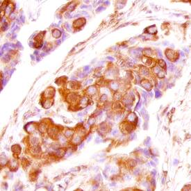
FGF‑5 in Human Breast Cancer Tissue.
FGF-5 was detected in immersion fixed paraffin-embedded sections of human breast cancer tissue using Mouse Anti-Human FGF-5 Monoclonal Antibody (Catalog # MAB2371) at 25 µg/mL overnight at 4 °C. Tissue was stained using the Anti-Mouse HRP-DAB Cell & Tissue Staining Kit (brown; Catalog # CTS002) and counterstained with hematoxylin (blue). Specific staining was localized to plasma membrane of ductal cells. View our protocol for Chromogenic IHC Staining of Paraffin-embedded Tissue Sections.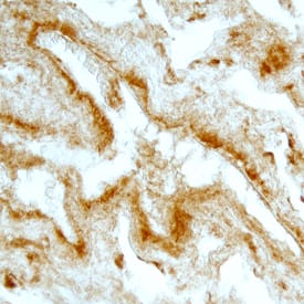
FGF‑5 in Human Breast Cancer Tissue.
FGF-5 was detected in immersion fixed paraffin-embedded sections of human breast cancer tissue using Mouse Anti-Human FGF-5 Monoclonal Antibody (Catalog # MAB2371) at 25 µg/mL overnight at 4 °C. Tissue was stained using the Anti-Mouse HRP-DAB Cell & Tissue Staining Kit (brown; Catalog # CTS002) and counterstained with hematoxylin (blue). Specific staining was localized to plasma membrane of ductal cells. View our protocol for Chromogenic IHC Staining of Paraffin-embedded Tissue Sections.
Detection of FGF-5 by Western Blot
The expression changes of ERK, AKT, p-CREB, c-fos, STAT3, PCNA, Cyclin A2, and Cyclin E after treatment of FGF5. a, b Western blot revealed the expression changes of p-ERK and p-AKT after 100-ng/ml FGF5 treatment. ERK, AKT, and ACTB were used as loading controls. c The expression changes of p-CREB, c-fos, and STAT3 after treatment of FGF5 for 5 and 15 min. d The expression changes of PCNA, Cyclin A2, and Cyclin E after 100 ng/ml FGF5 treatment for 6 and 12 h Image collected and cropped by CiteAb from the following open publication (https://pubmed.ncbi.nlm.nih.gov/30670081), licensed under a CC-BY license. Not internally tested by R&D Systems.Applications for Human FGF-5 Antibody
Application
Recommended Usage
Immunohistochemistry
8-25 µg/mL
Sample: Immersion fixed paraffin-embedded sections of human lung and human breast cancer tissue
Sample: Immersion fixed paraffin-embedded sections of human lung and human breast cancer tissue
Reviewed Applications
Read 1 review rated 5 using MAB2371 in the following applications:
Formulation, Preparation, and Storage
Purification
Protein A or G purified from hybridoma culture supernatant
Reconstitution
Reconstitute at 0.5 mg/mL in sterile PBS. For liquid material, refer to CoA for concentration.
Formulation
Lyophilized from a 0.2 μm filtered solution in PBS with Trehalose. *Small pack size (SP) is supplied either lyophilized or as a 0.2 µm filtered solution in PBS.
Shipping
Lyophilized product is shipped at ambient temperature. Liquid small pack size (-SP) is shipped with polar packs. Upon receipt, store immediately at the temperature recommended below.
Stability & Storage
Use a manual defrost freezer and avoid repeated freeze-thaw cycles.
- 12 months from date of receipt, -20 to -70 °C as supplied.
- 1 month, 2 to 8 °C under sterile conditions after reconstitution.
- 6 months, -20 to -70 °C under sterile conditions after reconstitution.
Background: FGF-5
The FGF family is comprised of at least seven polypeptides that are potent regulators of cell proliferation, differentiation and function. All FGFs have two conserved cysteine residues and share 30-50% sequence homology at the amino acid level. FGF-5 was originally identified as a transforming gene by the NIH-3T3 focus formation assay using DNA derived from human tumors. FGF-5 cDNA encodes a 267 amino acid residue protein with a putative 22 amino acid residue signal peptide. The murine homologue of FGF-5 was cloned and found to be 84% homologous to the human protein at the amino acid sequence level. Human and murine FGF-5 exhibit cross species activity.
In vitro, rhFGF-5 is a mitogen for Balb/3T3 fibroblasts and bovine heart endothelial cells. FGF-5 was also reported to be a major muscle-derived survival factor for cultured spinal motoneurons. In vivo, FGF-5 is suggested to play important roles in both embryology and neurobiology. Developmentally, FGF-5 mRNA is initially found in the embryoblast followed by the lateral somatic mesoderm, where it may play a role in angiogenesis, plus the myotomes cranial to the tail region, where it may delay terminal myoblast differentiation during cell migration. FGF-5 continues to impact muscle post-natally where it is believed to function as a target-derived neurotrophic factor of skeletal muscle. In the nervous system, FGF-5 has been most often identified in neurons associated with the limbic system, notably in neurons of the olfactory bulb and pyramidal cells of the hippocampus. Hippocampal FGF-5 is suggested to serve as a neurotrophic and differentiative factor for cholinergic and serotonergic neurons projecting to this region.
Long Name
Fibroblast Growth Factor 5
Alternate Names
FGF5
Entrez Gene IDs
2250 (Human)
Gene Symbol
FGF5
UniProt
Additional FGF-5 Products
Product Documents for Human FGF-5 Antibody
Product Specific Notices for Human FGF-5 Antibody
For research use only
Loading...
Loading...
Loading...
Loading...
Loading...