
Human FBXW7/Cdc4 Antibody
R&D Systems, part of Bio-Techne | Catalog # MAB7776

Scientific images may be resized, cropped, or adjusted for brightness or contrast for presentation purposes. These updates are limited to presentation and do not affect the underlying scientific interpretation of the data. Where available, additional source data may be provided upon request.
Conjugate
Catalog #
Key Product Details
Species Reactivity
Validated:
Human
Cited:
Human, Mouse
Applications
Validated:
Western Blot
Cited:
Western Blot, Immunocytochemistry
Label
Unconjugated
Antibody Source
Monoclonal Mouse IgG2B Clone # 800201
Product Specifications
Immunogen
E. coli-derived recombinant human FBXW7/Cdc4
Lys377-Lys707
Accession # Q969H0
Lys377-Lys707
Accession # Q969H0
Specificity
Detects human FBXW7/Cdc4 in direct ELISAs.
Clonality
Monoclonal
Host
Mouse
Isotype
IgG2B
Scientific Data Images for Human FBXW7/Cdc4 Antibody
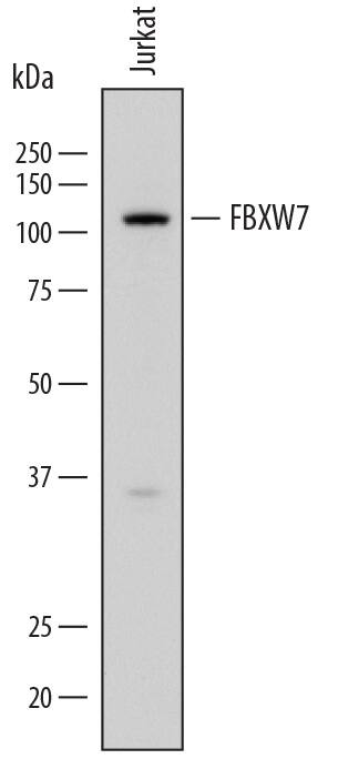
Detection of Human FBXW7/Cdc4 by Western Blot.
Western blot shows lysates of Jurkat human acute T cell leukemia cell line. PVDF membrane was probed with 2 µg/mL of Mouse Anti-Human FBXW7/Cdc4 Monoclonal Antibody (Catalog # MAB7776) followed by HRP-conjugated Anti-Mouse IgG Secondary Antibody (Catalog # HAF018). A specific band was detected for FBXW7/Cdc4 at approximately 110 kDa (as indicated). This experiment was conducted under reducing conditions and using Immunoblot Buffer Group 1.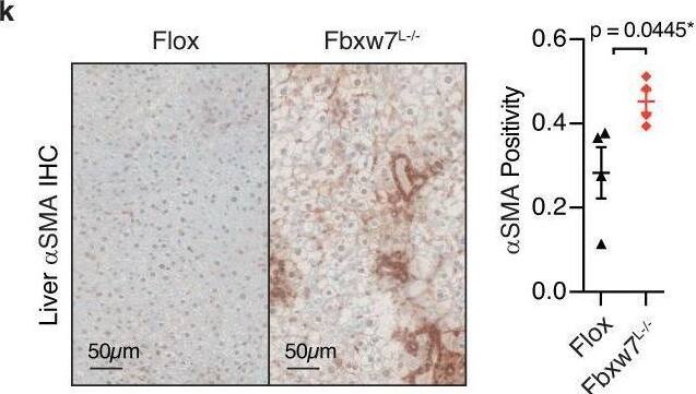
Detection of brqg9ujed6vbcpgfvygo.jpg by Immunohistochemistry
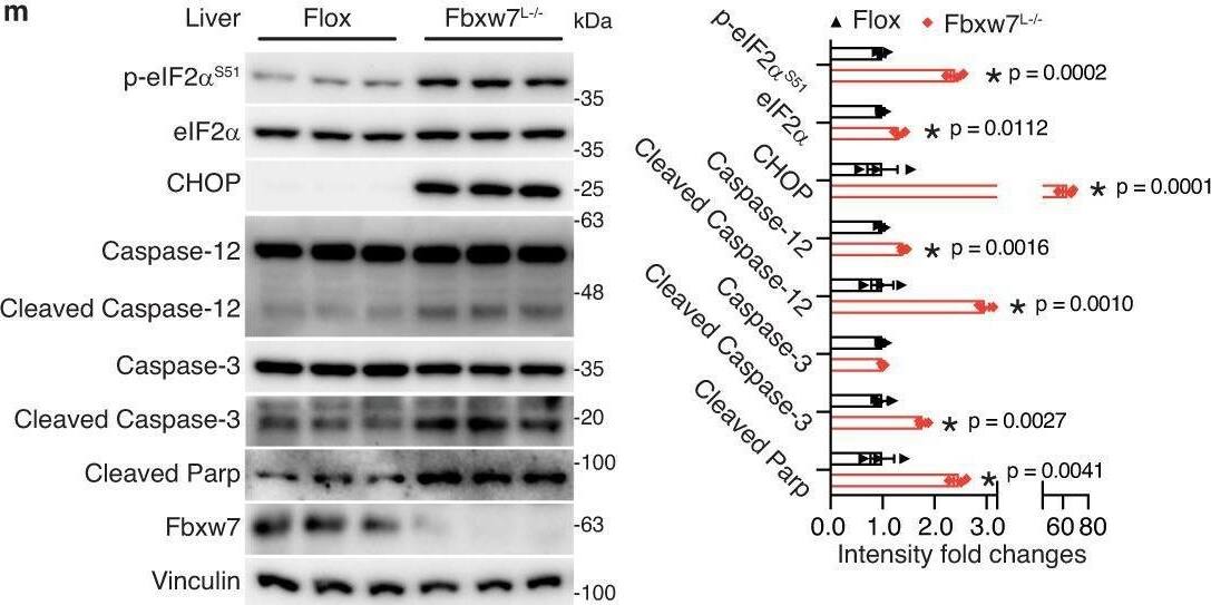
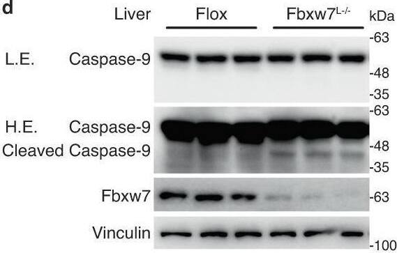
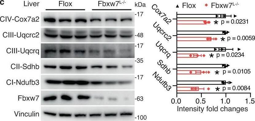
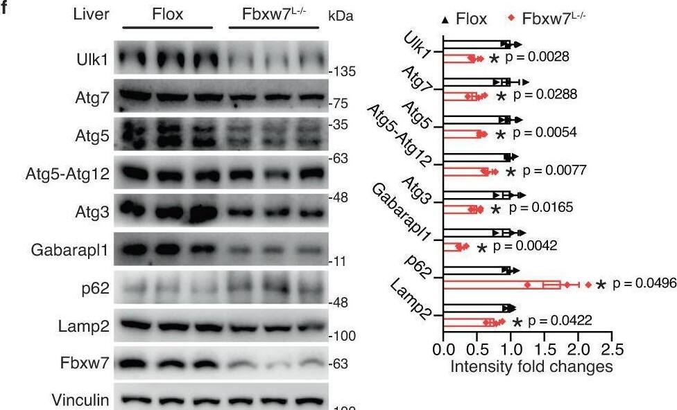
FBXW7 protects against NASH.a Analysis of hepatic FBXW7 expression in patients across the severity stages of NAFLD progression. n = 50 for NAFL; n = 87 for NASH_F0-2; n = 68 for NASH_F3-4. bFBXW7 mRNA levels in fibrotic liver biopsies from patients with NAFLD. n = 137 for Fibrosis stage 0-2; n = 68 for Fibrosis stage 3-4. Relative mRNA levels of NASH progression signature genes (c), inflammation and fibrogenesis genes (d) in Flox (control) and Fbxw7-null livers, n = 5. e Immunoblots and quantification of proteins related to NASH progression in livers and serum from Flox and Fbxw7L−/− littermates. Each lane represents one mouse, n = 3. f Overall immune contents of control and Fbxw7-null livers, reflected by the absolute immune fraction scores, n = 3. g Stacked bar chart illustrating cell distributions of control and Fbxw7-null livers, n = 3. CAFs Cancer-associated fibroblasts, na non-applicable. GSEA of SAMac-defining signature (h) and hepatic stellate cell signature (i). Array genes were ordered from the highest in Fbxw7-null livers (left) to the highest in control livers (right). Locations of genes in each signature are indicated by the vertical black bars. NES normalized enrichment score. Representative images and quantification of Aif1 (j) and alphaSMA (k) immunohistochemistry (IHC) staining, n = 4. Scale bars, 50 μm. l mRNA expression of ER stress and apoptotic genes in control and Fbxw7-null livers, n = 5. m Immunoblots and quantification of proteins related to ER stress and apoptosis. Each lane represents one mouse, n = 3. Data are presented as means ± SEM (a–f, j–m). *p < 0.05, unpaired two-tailed Student’s t test (a–g, j–m). Source data are provided as a Source Data file. Image collected and cropped by CiteAb from the following open publication (https://pubmed.ncbi.nlm.nih.gov/37914694), licensed under a CC-BY license. Not internally tested by R&D Systems.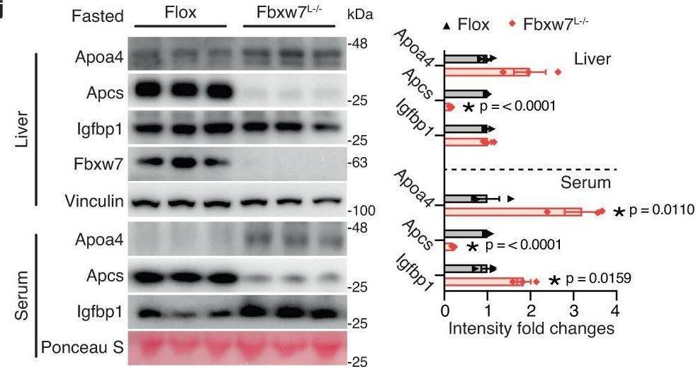
Detection of chnbnm42rnebvmhsgzk7.jpg by Western Blot
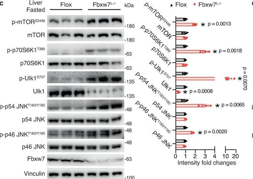
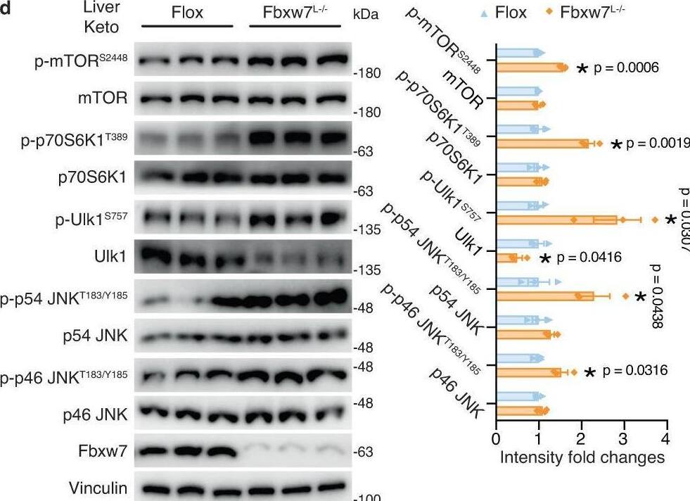
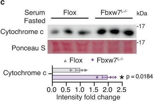
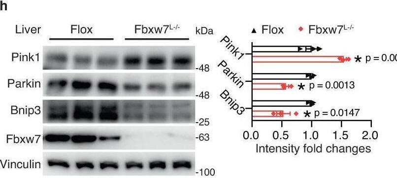
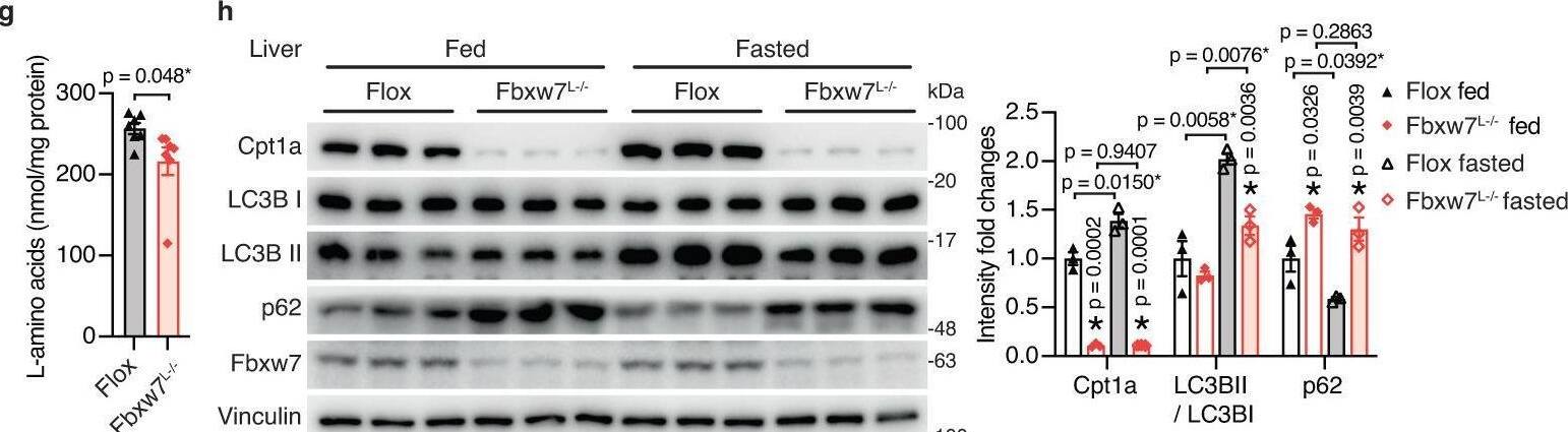
Metabolic stress aggravates NASH in the absence of Fbxw7.a Heatmap showing mRNA fold changes of NASH-related genes in the fed, fasted and keto diet nutritional regimens, assessed by RT-qPCR, n = 5-6 (see Source data file for specific sample numbers). b Representative images of H&E and Masson’s trichrome (MTC) stained liver tissue sections from Flox and Fbxw7L−/− mice under different nutritional regimens. Arrowhead, fibrotic area (blue). Images are representative of n = 3 mice per group. Immunoblots and quantification of the indicated proteins from Flox and Fbxw7L−/− mice upon fasting (c) or post a 20-day keto diet (d). Each lane represents one mouse, n = 3. e HepG2 cells stably expressing empty vector or FBXW7 were treated with 500 nM Thapsigargin (Tg) or DMSO for 24 h followed by immunoblot examination. Immunoblots are from one experiment representative of 3 independent experiments with similar results. f Overlap of liver-enriched secreted factors with hepatic DEGs (p < 0.05, |FC|>1.30) induced by Fbxw7 deficiency, by fasting, or by a keto diet. g Ranking of the secreted factors identified in Fig. 7f by the sum of absolute mRNA fold changes of the four transcriptome datasets. h, i Hepatic mRNA expression of Apoa4, Apcs and Igfbp1 from Flox and Fbxw7L−/− mice upon fasting (h, n = 5) or post a 20-day keto diet (i, n = 6 for Flox and n = 5 for Fbxw7L−/− mice). Immunoblots and quantification of serum and liver levels of the hepatic secreted factors from Flox and Fbxw7L−/− mice upon fasting (j) or post a 20-day keto diet (k). Each lane represents one mouse, n = 3. Data are represented as means ± SEM (c, d, h–k). *p < 0.05, unpaired two-tailed Student’s t test (a, c, d, h–k). Source data are provided as a Source Data file including Fig. 7a p-values. Image collected and cropped by CiteAb from the following open publication (https://pubmed.ncbi.nlm.nih.gov/37914694), licensed under a CC-BY license. Not internally tested by R&D Systems.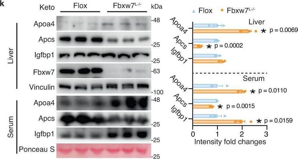
Applications for Human FBXW7/Cdc4 Antibody
Application
Recommended Usage
Western Blot
2 µg/mL
Sample: Jurkat human acute T cell leukemia cell line
Sample: Jurkat human acute T cell leukemia cell line
Reviewed Applications
Read 3 reviews rated 4 using MAB7776 in the following applications:
Formulation, Preparation, and Storage
Purification
Protein A or G purified from hybridoma culture supernatant
Reconstitution
Sterile PBS to a final concentration of 0.5 mg/mL. For liquid material, refer to CoA for concentration.
Formulation
Lyophilized from a 0.2 μm filtered solution in PBS with Trehalose. *Small pack size (SP) is supplied either lyophilized or as a 0.2 µm filtered solution in PBS.
Shipping
Lyophilized product is shipped at ambient temperature. Liquid small pack size (-SP) is shipped with polar packs. Upon receipt, store immediately at the temperature recommended below.
Stability & Storage
Use a manual defrost freezer and avoid repeated freeze-thaw cycles.
- 12 months from date of receipt, -20 to -70 °C as supplied.
- 1 month, 2 to 8 °C under sterile conditions after reconstitution.
- 6 months, -20 to -70 °C under sterile conditions after reconstitution.
Background: FBXW7/Cdc4
Long Name
F-box and WD Repeat Domain Containing 7
Alternate Names
AGO, Archipelago Homolog, Cdc4, FBW6, Fbw7, FBX30, FBXO30, FBXW6, SCF(Fbw7), SEL10
Gene Symbol
FBXW7
UniProt
Additional FBXW7/Cdc4 Products
Product Documents for Human FBXW7/Cdc4 Antibody
Product Specific Notices for Human FBXW7/Cdc4 Antibody
For research use only
Loading...
Loading...
Loading...
Loading...
Loading...