
Human EGFR PE-conjugated Antibody
R&D Systems, part of Bio-Techne | Catalog # FAB10951P

Scientific images may be resized, cropped, or adjusted for brightness or contrast for presentation purposes. These updates are limited to presentation and do not affect the underlying scientific interpretation of the data. Where available, additional source data may be provided upon request.
Conjugate
Catalog #
Key Product Details
Species Reactivity
Validated:
Human
Cited:
Human
Applications
Validated:
Flow Cytometry
Cited:
Flow Cytometry, Immunocytochemistry
Label
Phycoerythrin (Excitation = 488 nm, Emission = 565-605 nm)
Antibody Source
Monoclonal Rat IgG2A Clone # 423103
Product Specifications
Immunogen
Mouse myeloma cell line NS0-derived recombinant human EGF R/ErbB1
Leu25-Ser645
Accession # CAA25240
Leu25-Ser645
Accession # CAA25240
Specificity
Detects human EGF R/ErbB1 in direct ELISAs. In direct ELISAs, no cross-reactivity with recombinant human (rh) ErbB2, rhErbB3, or rhErbB4 is observed.
Clonality
Monoclonal
Host
Rat
Isotype
IgG2A
Scientific Data Images for Human EGFR PE-conjugated Antibody
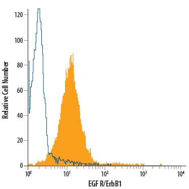
Detection of EGF R/ErbB1 in A431 human epithelial carcinoma cell line by Flow Cytometry.
A431 human epithelial carcinoma cell line was stained with Rat Anti-Human EGF R/ErbB1 PE-conjugated Monoclonal Antibody (Catalog # FAB10951P, filled histogram) or isotype control antibody (Catalog # IC006P, open histogram). View our protocol for Staining Membrane-associated Proteins.Applications for Human EGFR PE-conjugated Antibody
Application
Recommended Usage
Flow Cytometry
10 µL/106 cells
Sample: A431 human epithelial carcinoma cell line
Sample: A431 human epithelial carcinoma cell line
Spectra Viewer
Plan Your Experiments
Use our spectra viewer to interactively plan your experiments, assessing multiplexing options. View the excitation and emission spectra for our fluorescent dye range and other commonly used dyes.
Spectra Viewer
Formulation, Preparation, and Storage
Purification
Protein A or G purified from hybridoma culture supernatant
Formulation
Supplied in a saline solution containing BSA and Sodium Azide.
Shipping
The product is shipped with polar packs. Upon receipt, store it immediately at the temperature recommended below.
Stability & Storage
Protect from light. Do not freeze.
- 12 months from date of receipt, 2 to 8 °C as supplied.
Background: EGFR
Epidermal Growth Factor Receptor (EGF R), also named erythroblastic leukemia viral oncogene homolog 1 (ErbB1), is a member of the type I receptor tyrosine kinase superfamily. The epidermal growth factor receptor (EGF R) subfamily of receptor tyrosine kinases comprises four members: EGF R (also known as HER1, ErbB1or ErbB), ErbB2 (Neu, HER2), ErbB3 (HER3), and ErbB4 (HER4). All family members are type I transmembrane glycoproteins that have an extracellular domain with two ligand binding cysteine rich domains, separated by a spacer region, and a cytoplasmic domain with a membrane proximal tyrosine kinase domain and a C-terminal tail with multiple tyrosine autophosphorylation sites. The human EGF R geneencodes a 1210 amino acid (aa) residue precursor with a 24 aa putative signal peptide, a 621 aa extracellular domain, a 23 aa transmembrane domain, and a 542 aa cytoplasmic domain. EGF R has been shown to bind a subset of the EGF family ligands, including EGF, amphiregulin, TGF alpha, betacellulin, epiregulin, heparin-binding EGF and neuregulin-2 alpha, in the absence of a coreceptor. Ligand binding induces EGF R homodimerization as well as heterodimerization with ErbB2, resulting in kinase activation, tyrosine phosphorylation and cell signaling. EGF R can also be recruited to form heterodimers with ligand-activated ErbB3 or ErbB4. EGF R signaling has been shown to regulate multiple biological functions including cell proliferation, differentiation, motility and apoptosis. In addition, EGF R signaling has also been shown to play a role in carcinogenesis (1 ‑ 3).
References
- Daly, R.J. (1999) Growth Factors, 16:255.
- Schlessinger, J. (2000) Cell. 103:211.
- Maihle, N.J. et al. (2002) Cancer Treat. Res. 107:247.
Long Name
Epidermal Growth Factor Receptor
Alternate Names
EGF R, ErbB, ErbB1, HER-1
Gene Symbol
EGFR
UniProt
Additional EGFR Products
Product Documents for Human EGFR PE-conjugated Antibody
Product Specific Notices for Human EGFR PE-conjugated Antibody
For research use only
Loading...
Loading...
Loading...
Loading...
Loading...