
Human DcR3/TNFRSF6B Antibody
R&D Systems, part of Bio-Techne | Catalog # AF142


Scientific images may be resized, cropped, or adjusted for brightness or contrast for presentation purposes. These updates are limited to presentation and do not affect the underlying scientific interpretation of the data. Where available, additional source data may be provided upon request.
Conjugate
Catalog #
Key Product Details
Species Reactivity
Validated:
Human
Cited:
Human
Applications
Validated:
Immunohistochemistry, Western Blot, Neutralization
Cited:
Flow Cytometry
Label
Unconjugated
Antibody Source
Polyclonal Goat IgG
Product Specifications
Immunogen
S. frugiperda insect ovarian cell line Sf 21-derived recombinant human DcR3/TNFRSF6B
Val24-His300
Accession # O95407
Val24-His300
Accession # O95407
Specificity
Detects human DcR3/TNFRSF6B in ELISAs and Western blots. In direct ELISAs, less than 1% cross-reactivity with recombinant human (rh) TRAIL R1, rhTRAIL R2, rhTRAIL R3, and rhTRAIL R4 is observed.
Clonality
Polyclonal
Host
Goat
Isotype
IgG
Endotoxin Level
<0.10 EU per 1 μg of the antibody by the LAL method.
Scientific Data Images for Human DcR3/TNFRSF6B Antibody
DcR3/TNFRSF6B in Human Stomach Cancer Tissue.
DcR3/TNFRSF6B was detected in immersion fixed paraffin-embedded sections of human stomach cancer using 10 µg/mL Goat Anti-Human DcR3/TNFRSF6B Antigen Affinity-purified Polyclonal Antibody (Catalog # AF142) overnight at 4 °C. Before incubation with the primary antibody tissue was subjected to heat-induced epitope retrieval using Antigen Retrieval Reagent-Basic (Catalog # CTS013). Tissue was stained with the Anti-Goat HRP-DAB Cell & Tissue Staining Kit (brown; Catalog # CTS008) and counterstained with hematoxylin (blue). Specific labeling was localized to the membrane of epithelial cells in gastric glands. View our protocol for Chromogenic IHC Staining of Paraffin-embedded Tissue Sections.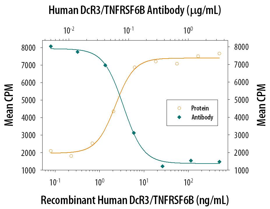
DcR3/TNFRSF6B Inhibition of Fas Ligand/TNFSF6-induced Apoptosis and Neutralization by Human DcR3/TNFRSF6B Antibody.
In the presence of a cross-linking antibody, Mouse polyHistidine Monoclonal Antibody (10 µg/mL, Catalog # MAB050), Recombinant Human DcR3/TNFRSF6B Fc Chimera (Catalog # 142-DC) inhibits Recombinant Human Fas Ligand/TNFSF6 (Catalog # 126-FL) induced apoptosis in the Jurkat human acute T cell leukemia cell line in a dose-dependent manner (orange line). Under these conditions, inhibition of Recombinant Human Fas Ligand/TNFSF6 (2 ng/mL) activity elicited by Recombinant Human DcR3/TNFRSF6B Fc Chimera (50 ng/mL) is neutralized (green line) by increasing concentrations of Goat Anti-Human DcR3/TNFRSF6B Antigen Affinity-purified Polyclonal Antibody (Catalog # AF142). The ND50 is typically 0.1-0.5 µg/mL.Applications for Human DcR3/TNFRSF6B Antibody
Application
Recommended Usage
Immunohistochemistry
5-15 µg/mL
Sample: Immersion fixed paraffin-embedded sections of human stomach cancer subjected to Antigen Retrieval Reagent-Basic (Catalog # CTS013)
Sample: Immersion fixed paraffin-embedded sections of human stomach cancer subjected to Antigen Retrieval Reagent-Basic (Catalog # CTS013)
Western Blot
0.1 µg/mL
Sample: Recombinant Human DcR3/TNFRSF6B Fc Chimera (Catalog # 142-DC)
Sample: Recombinant Human DcR3/TNFRSF6B Fc Chimera (Catalog # 142-DC)
Neutralization
Measured by its ability to neutralize DcR3/TNFRSF6B-mediated inhibition of apoptosis in the Jurkat human acute T cell leukemia cell line. The Neutralization Dose (ND50) is typically 0.1-0.5 µg/mL in the presence of 50 ng/mL Recombinant Human DcR3/TNFRSF6B Fc Chimera, 2 ng/mL Recombinant Human Fas Ligand/TNFSF6.
Formulation, Preparation, and Storage
Purification
Antigen Affinity-purified
Reconstitution
Reconstitute at 0.2 mg/mL in sterile PBS. For liquid material, refer to CoA for concentration.
Formulation
Lyophilized from a 0.2 μm filtered solution in PBS with Trehalose. *Small pack size (SP) is supplied either lyophilized or as a 0.2 µm filtered solution in PBS.
Shipping
Lyophilized product is shipped at ambient temperature. Liquid small pack size (-SP) is shipped with polar packs. Upon receipt, store immediately at the temperature recommended below.
Stability & Storage
Use a manual defrost freezer and avoid repeated freeze-thaw cycles.
- 12 months from date of receipt, -20 to -70 °C as supplied.
- 1 month, 2 to 8 °C under sterile conditions after reconstitution.
- 6 months, -20 to -70 °C under sterile conditions after reconstitution.
Background: DcR3/TNFRSF6B
Human decoy receptor 3 (DcR3), also called TNFRSF6B, TR6, and M68, is a member of the TNF receptor superfamily. The cDNA of DcR3 encodes a 300 amino acid (aa) polypeptide with a putative 23 aa signal peptide. Like osteoprotegerin (OPG), DcR3 lacks a transmembrane sequence and is a secreted protein. DcR3 shares sequence identity with OPG (31%), TNF RII (29%), and Fas (17%). It was found to be expressed in a variety of different tissues and at high levels in many malignant tumors. Among TNF family members, DcR3 was shown to bind with Fas ligand (FasL) and LIGHT and inhibit FasL- and LIGHT-induced apoptosis. Thus, in addition to DcR1, DcR2, and OPG, DcR3 is another TNFR family molecule which modulates ligands that induce apoptosis. Over-expression of DcR3 might be a mechanism by which certain tumors escape immune-cytotoxic attack.
References
- Pitti, R.M. et al. (1998) Nature 396:699.
- Yu, K-Y. et al. (1999) J. Biochem. Chem. 274:13733.
- Bai, C. et al. (2000) Proc. Natl. Acad. Sci. USA 97:1230.
Long Name
Decoy Receptor 3
Alternate Names
M68, TNFRSF6B, TR6
Gene Symbol
TNFRSF6B
UniProt
Additional DcR3/TNFRSF6B Products
Product Documents for Human DcR3/TNFRSF6B Antibody
Product Specific Notices for Human DcR3/TNFRSF6B Antibody
For research use only
Loading...
Loading...
Loading...
Loading...
Loading...