
Human CD40 Ligand/TNFSF5 Antibody
R&D Systems, part of Bio-Techne | Catalog # AF617

Scientific images may be resized, cropped, or adjusted for brightness or contrast for presentation purposes. These updates are limited to presentation and do not affect the underlying scientific interpretation of the data. Where available, additional source data may be provided upon request.
Conjugate
Catalog #
Key Product Details
Species Reactivity
Validated:
Human
Cited:
Human
Applications
Validated:
Western Blot, Flow Cytometry, CyTOF-ready
Cited:
Immunohistochemistry-Frozen, Western Blot, Flow Cytometry, Dot Blot
Label
Unconjugated
Antibody Source
Polyclonal Goat IgG
Product Specifications
Immunogen
E. coli-derived recombinant human CD40 Ligand/TNFSF5
Glu108-Leu261
Accession # P29965
Glu108-Leu261
Accession # P29965
Specificity
Detects human CD40 Ligand/TNFSF5 in direct ELISAs and Western blots. In direct ELISAs, approximately 20% cross-reactivity with recombinant mouse CD40 Ligand is observed and less than 1% cross-reactivity with recombinant human (rh) CD27 Ligand and rhCD30 Ligand is observed.
Clonality
Polyclonal
Host
Goat
Isotype
IgG
Scientific Data Images for Human CD40 Ligand/TNFSF5 Antibody
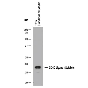
Detection of Human CD40 Ligand/TNFSF5 by Western Blot.
Western blot shows conditioned media (CM) from Th17 cells. PVDF membrane was probed with 1 µg/mL of Goat Anti-Human CD40 Ligand/TNFSF5 Antigen Affinity-purified Polyclonal Antibody (Catalog # AF617) followed by HRP-conjugated Anti-Goat IgG Secondary Antibody (Catalog # HAF017). A specific band was detected for CD40 Ligand/TNFSF5 at approximately 17 kDa (as indicated). This experiment was conducted under reducing conditions and using Western Blot Buffer Group 1.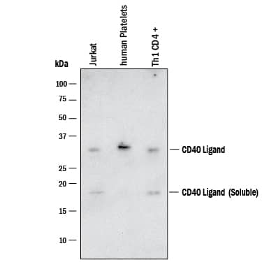
Detection of Human CD40 Ligand/TNFSF5 by Western Blot.
Western blot shows lysates of Jurkat human acute T cell leukemia cells, Human Platelets and Th1 CD4+. PVDF membrane was probed with 1 µg/mL of Goat Anti-Human CD40 Ligand/TNFSF5 Antigen Affinity-purified Polyclonal Antibody (Catalog # AF617) followed by HRP-conjugated Anti-Goat IgG Secondary Antibody (Catalog # HAF017). Specific bands were detected for CD40 Ligand/TNFSF5 at approximately 17 and 32 kDa (as indicated). This experiment was conducted under reducing conditions and using Western Blot Buffer Group 1.
Detection of CD40 Ligand/TNFSF5 by Flow Cytometry
Characterization of SHIV DNA. (A) Schematic of SHIV DNA construct. (B) Western blot analysis of SIV Gag, HIV C.1086 Env, and Rhesus CD40L in the lysate and supernatant of transfected 293 T cells. (C) Flow cytometry of 293 transfected cells depicting the surface co-expression of HIV Env and rhesus CD40L on SIV Gag positive cells (D) Surface binding of bNAbs and non-bNAbs to SIV Gag positive DNA transfected cells. Image collected and cropped by CiteAb from the following open publication (https://www.frontiersin.org/articles/10.3389/fimmu.2022.914969/full), licensed under a CC-BY license. Not internally tested by R&D Systems.Applications for Human CD40 Ligand/TNFSF5 Antibody
Application
Recommended Usage
CyTOF-ready
Ready to be labeled using established conjugation methods. No BSA or other carrier proteins that could interfere with conjugation.
Flow Cytometry
2.5 µg/106 cells
Sample: Human peripheral blood mononuclear cells treated with PMA and Ca2+ ionomycin
Sample: Human peripheral blood mononuclear cells treated with PMA and Ca2+ ionomycin
Western Blot
1 µg/mL
Sample: Th17 cells, Jurkat human acute T cell leukemia cells, Human Platelets and Th1 CD4+
Sample: Th17 cells, Jurkat human acute T cell leukemia cells, Human Platelets and Th1 CD4+
Formulation, Preparation, and Storage
Purification
Antigen Affinity-purified
Reconstitution
Reconstitute at 0.2 mg/mL in sterile PBS. For liquid material, refer to CoA for concentration.
Formulation
Lyophilized from a 0.2 μm filtered solution in PBS with Trehalose. *Small pack size (SP) is supplied either lyophilized or as a 0.2 µm filtered solution in PBS.
Shipping
Lyophilized product is shipped at ambient temperature. Liquid small pack size (-SP) is shipped with polar packs. Upon receipt, store immediately at the temperature recommended below.
Stability & Storage
Use a manual defrost freezer and avoid repeated freeze-thaw cycles.
- 12 months from date of receipt, -20 to -70 °C as supplied.
- 1 month, 2 to 8 °C under sterile conditions after reconstitution.
- 6 months, -20 to -70 °C under sterile conditions after reconstitution.
Background: CD40 Ligand/TNFSF5
CD40 ligand, CD40L (also known as CD154, TRAP or gp39), is a 261 amino acid type II transmembrane glycoprotein belonging to the TNF family, CD40L is expressed predominantly on activated CD4+ T lymphocytes, and also found in other types of cells, like NK cells, mast cells, basophils and eosinophils. Human CD40L shares 78% amino acid identity with its murine counterpart. The receptor of CD40L is CD40, a type I transmembrane glycoprotein belonging to the TNF receptor family. CD40 is expressed on B lymphocytes, monocytes, dendritic cells and thymic epithelium. Although all monomeric, dimeric and trimeric forms of soluble CD40L can bind to CD40, the trimeric form of soluble CD40L has the most potent biological activity through oligomerization of cell surface CD40, a common feature of TNF receptor family members. CD40L mediates a range of activities on B cells including induction of activation-associated surface antigen, entry into cell cycle, isotype switching and Ig secretion and memory generation. CD40-CD40L interaction also plays important roles in monocyte activation and dendritic cell maturation.
References
- Armitage, R.J. et al. (1992) Nature 357:80.
- Hollenbaugh, D. et al. (1992) EMBO J. 11:4313.
- Spriggs, M.K. et al. (1992) J. Exp. Med. 176:1543.
- Fanslow, W.C. et al. (1994) Seminars in Immunology 6:267.
- Kooten, C.V. and J. Banchereau (2000) J. Leukoc. Biol. 67:2.
Alternate Names
CD154, CD40L, CD40LG, gp39, TNFSF5, TRAP
Gene Symbol
CD40LG
UniProt
Additional CD40 Ligand/TNFSF5 Products
Product Documents for Human CD40 Ligand/TNFSF5 Antibody
Product Specific Notices for Human CD40 Ligand/TNFSF5 Antibody
For research use only
Loading...
Loading...
Loading...
Loading...
Loading...
Loading...