
Human CD30/TNFRSF8 Antibody
R&D Systems, part of Bio-Techne | Catalog # AF229

Scientific images may be resized, cropped, or adjusted for brightness or contrast for presentation purposes. These updates are limited to presentation and do not affect the underlying scientific interpretation of the data. Where available, additional source data may be provided upon request.
Conjugate
Catalog #
Key Product Details
Species Reactivity
Human
Applications
Western Blot, ELISA, Immunocytochemistry, Simple Western, Agonist Activity
Label
Unconjugated
Antibody Source
Polyclonal Goat IgG
Product Specifications
Immunogen
S. frugiperda insect ovarian cell line Sf 21-derived recombinant human CD30
Phe19-Lys379
Accession # P28908
Phe19-Lys379
Accession # P28908
Specificity
Detects human CD30 in direct ELISAs and Western blots.
Clonality
Polyclonal
Host
Goat
Isotype
IgG
Endotoxin Level
<0.10 EU per 1 μg of the antibody by the LAL method.
Scientific Data Images for Human CD30/TNFRSF8 Antibody
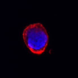
CD30/TNFRSF8 in Jurkat Human Cell Line.
CD30/TNFRSF8 was detected in immersion fixed Jurkat human acute T cell leukemia cell line using Goat Anti-Human CD30/TNFRSF8 Antigen Affinity-purified Polyclonal Antibody (Catalog # AF229) at 10 µg/mL for 3 hours at room temperature. Cells were stained using the NorthernLights™ 557-conjugated Anti-Goat IgG Secondary Antibody (red; NL001) and counterstained with DAPI (blue). Specific staining was localized to cell membrane and cytoplasm. View our protocol for Fluorescent ICC Staining of Non-adherent Cells.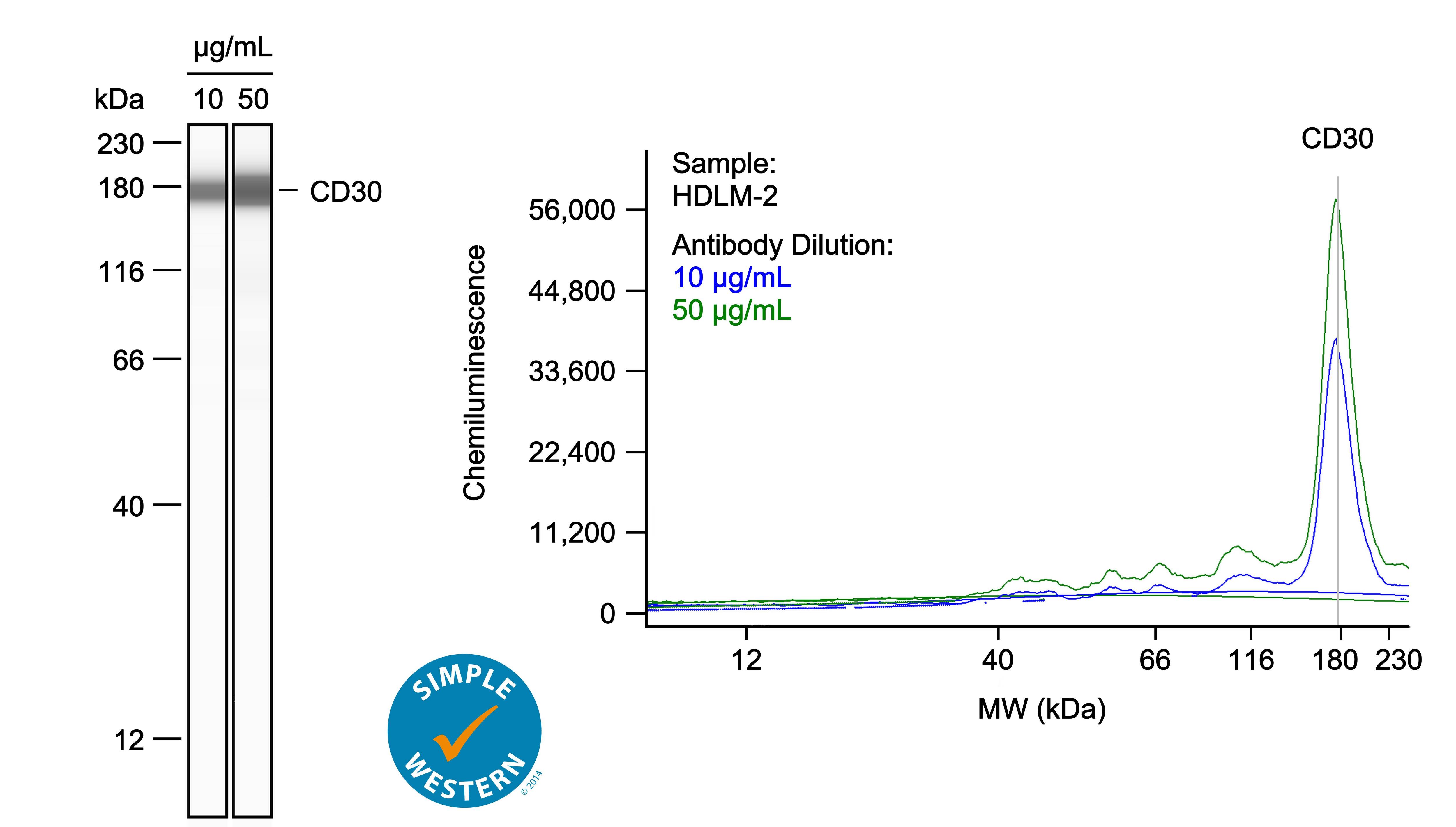
Detection of Human CD30/TNFRSF8 by Simple WesternTM.
Left: Simple Western lane view shows lysates of HDLM-2 human Hodgkin’s lymphoma cell line, loaded at 0.5 mg/ml. A specific band was detected for CD30/TNFRSF8 at approximately 178 kDa (as indicated) using both 10 µg/ml and 50 µg/ml of Goat Anti-Human CD30/TNFRSF8 Antigen Affinity-purified Polyclonal Antibody (Catalog # AF229) followed by HRP-conjugated Donkey Anti-Goat Secondary Antibody (Catalog # 043-522-2). This experiment was conducted under reducing conditions and using the 12-230kDa separation system. Right: Simple Western electropherogram showing the same Goat Anti-Human CD30/TNFRSF8 Antigen Affinity-purified Polyclonal Antibody (Catalog # AF229) tested at 10 µg/ml (blue line) and 50 µg/ml (green line) in the HDLM-2 human Hodgkin’s lymphoma cell line.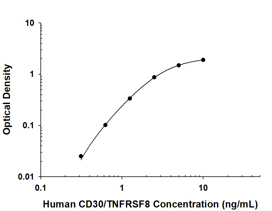
Human CD30/TNFRSF8 ELISA Standard Curve.
Recombinant Human CD30/TNFRSF8 protein was serially diluted 2-fold and captured by Mouse Anti-Human CD30/TNFRSF8 Monoclonal Antibody (MAB2291) coated on a Clear Polystyrene Microplate (DY990). Goat Anti-Human CD30/TNFRSF8 Antigen Affinity-purified Polyclonal Antibody (Catalog # AF229) was biotinylated and incubated with the protein captured on the plate. Detection of the standard curve was achieved by incubating Streptavidin-HRP (DY998) followed by Substrate Solution (DY999) and stopping the enzymatic reaction with Stop Solution (DY994).Applications for Human CD30/TNFRSF8 Antibody
Application
Recommended Usage
Agonist Activity
Measured by its ability to stimulate human IL-6 secretion by HDLM-2 human Hodgkin’s lymphoma cells. Gruss H.J. et al. (1996) Blood 87(6):2443.
The ED50 for this effect is typically 0.2 - 0.6 μg/mL.
The ED50 for this effect is typically 0.2 - 0.6 μg/mL.
ELISA
This antibody functions as an ELISA detection antibody when paired with Mouse Anti-Human CD30/TNFRSF8 Monoclonal Antibody (Catalog # MAB2291).
This product is intended for assay development on various assay platforms requiring antibody pairs. We recommend the Human CD30/TNFRSF8 DuoSet ELISA Kit (Catalog # DY6126-05) for convenient development of a sandwich ELISA.
Immunocytochemistry
5-15 µg/mL
Sample: Immersion fixed Jurkat human acute T cell leukemia cell line
Sample: Immersion fixed Jurkat human acute T cell leukemia cell line
Simple Western
10-50 µg/mL
Sample: HDLM-2 human Hodgkin's lymphoma cell line
Sample: HDLM-2 human Hodgkin's lymphoma cell line
Western Blot
0.1 µg/mL
Sample: Recombinant Human CD30/TNFRSF8 Fc Chimera (Catalog # 813-CD)
Sample: Recombinant Human CD30/TNFRSF8 Fc Chimera (Catalog # 813-CD)
Reviewed Applications
Read 1 review rated 5 using AF229 in the following applications:
Formulation, Preparation, and Storage
Purification
Antigen Affinity-purified
Reconstitution
Reconstitute at 0.2 mg/mL in sterile PBS. For liquid material, refer to CoA for concentration.
Formulation
Lyophilized from a 0.2 μm filtered solution in PBS with Trehalose. See Certificate of Analysis for details.
*Small pack size (-SP) is supplied either lyophilized or as a 0.2 µm filtered solution in PBS.
*Small pack size (-SP) is supplied either lyophilized or as a 0.2 µm filtered solution in PBS.
Shipping
Lyophilized product is shipped at ambient temperature. Liquid small pack size (-SP) is shipped with polar packs. Upon receipt, store immediately at the temperature recommended below.
Stability & Storage
Use a manual defrost freezer and avoid repeated freeze-thaw cycles.
- 12 months from date of receipt, -20 to -70 °C as supplied.
- 1 month, 2 to 8 °C under sterile conditions after reconstitution.
- 6 months, -20 to -70 °C under sterile conditions after reconstitution.
Background: CD30/TNFRSF8
References
- Kennedy, M.K. et al. (2006) Immunology 118:143.
- Tarkowski, M. (2003) Curr. Opin. Hematol. 10:267.
- Durkop, H. et al. (1992) Cell 68:421.
- Hamann, D. et al. (1996) J. Immunol. 156:1387.
- Shanebeck, S.D. et al. (1995) Eur. J. Immunol. 25:2147.
- Gruss, H.-J. et al. (1994) Blood 83:2045.
- Oflzoglu E. et al. (2009) Adv. Exp. Med. Biol. 647:174.
- Del Prete, G. et al. (1995) J. Exp. Med. 182:1655.
- Harlin, H. et al. (2002) J. Immunol. 169:2451.
- Amakawa, R. et al. (1996) Cell 84:551.
- Chiarle, R. et al. (1999) J. Immunol. 163:194.
- Vinante, F. et al. (2002) Blood 99:52.
- Hansen, H.P. et al. (1995) Int. J. Cancer 63:750.
- Hansen, H.P. et al. (2000) J. Immunol. 165:6703.
- Hargreaves, P.G. and A. Al-Shamkhani (2002) Eur. J. Immunol. 32:163.
Alternate Names
CD30, TNFRSF8
Gene Symbol
TNFRSF8
UniProt
Additional CD30/TNFRSF8 Products
Product Documents for Human CD30/TNFRSF8 Antibody
Product Specific Notices for Human CD30/TNFRSF8 Antibody
For research use only
Loading...
Loading...
Loading...
Loading...
Loading...