
Human CCL3/MIP-1 alpha Biotinylated Antibody Best Seller
R&D Systems, part of Bio-Techne | Catalog # BAF270

Scientific images may be resized, cropped, or adjusted for brightness or contrast for presentation purposes. These updates are limited to presentation and do not affect the underlying scientific interpretation of the data. Where available, additional source data may be provided upon request.
Conjugate
Catalog #
Key Product Details
Species Reactivity
Validated:
Human
Cited:
Human, Primate - Macaca fascicularis (Crab-eating Monkey or Cynomolgus Macaque)
Applications
Validated:
Western Blot, ELISA Detection (Matched Antibody Pair), Immunocytochemistry
Cited:
Immunohistochemistry, Immunohistochemistry-Paraffin, Western Blot, Immunocytochemistry, Antibody Array Development, ELISA Detection, ELISA Development, ELISpot Development, Luminex Development
Label
Biotin
Antibody Source
Polyclonal Goat IgG
Product Specifications
Immunogen
E. coli-derived recombinant human CCL3/MIP-1 alpha
Ala27-Ala92
Accession # P10147
Ala27-Ala92
Accession # P10147
Specificity
Detects human CCL3/MIP-1 alpha in ELISAs and Western blots. In sandwich immunoassays, less than 0.05% cross-reactivity with recombinant human (rh) MIP-1 beta, rhMIP-1 delta, rhMIP-3 alpha, rhMIP-3 beta, and recombinant mouse MIP-1 alpha is observed.
Clonality
Polyclonal
Host
Goat
Isotype
IgG
Scientific Data Images for Human CCL3/MIP-1 alpha Biotinylated Antibody
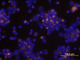
CCL3/MIP‑1 alpha in Human PBMCs.
CCL3/MIP-1a was detected in immersion fixed human peripheral blood mononuclear cells (PBMCs) stimulated with PHA and monensin using Human CCL3/MIP-1a Biotinylated Antigen Affinity-purified Polyclonal Antibody (Catalog # BAF270) at 10 µg/mL for 3 hours at room temperature. Cells were stained using the Northern-Lights™ 557-conjugated Streptavidin (yellow; Catalog # NL999) and counterstained with DAPI (blue). View our protocol for Fluorescent ICC Staining of Non-adherent Cells.
Human CCL3 / MIP-1 alpha ELISA Standard Curve
Recombinant Human CCL3/MIP-1 alpha Isoform LD78a (Catalog # 270-LD) was serially diluted and captured by Goat Anti-Human CCL3/MIP-1 alpha Antigen Affinity-purified Polyclonal Antibody (Catalog # AF-270-NA) coated on a Clear Polystyrene Microplate (Catalog # DY990). Goat Anti-Human CCL3/MIP-1 alpha Antigen Affinity-purified Polyclonal Antibody (Catalog # AF-270-NA) was biotinylated and incubated with the protein captured on the plate. Detection of the standard curve was achieved by incubating Streptavidin-HRP (Catalog # DY998)Applications for Human CCL3/MIP-1 alpha Biotinylated Antibody
Application
Recommended Usage
Immunocytochemistry
5-15 µg/mL
Sample: Immersion fixed human peripheral blood mononuclear cells (PBMCs) stimulated by PHA and monensin
Sample: Immersion fixed human peripheral blood mononuclear cells (PBMCs) stimulated by PHA and monensin
Western Blot
0.1 µg/mL
Sample: Recombinant Human CCL3/MIP-1 alpha isoform LD78a (Catalog # 270-LD)
Sample: Recombinant Human CCL3/MIP-1 alpha isoform LD78a (Catalog # 270-LD)
Human CCL3/MIP-1 alpha Sandwich Immunoassay
Please Note: Optimal dilutions of this antibody should be experimentally determined.
Formulation, Preparation, and Storage
Purification
Antigen Affinity-purified
Reconstitution
Reconstitute at 0.2 mg/mL in sterile PBS.
Formulation
Lyophilized from a 0.2 μm filtered solution in PBS with BSA as a carrier protein.
Shipping
The product is shipped at ambient temperature. Upon receipt, store it immediately at the temperature recommended below.
Stability & Storage
Use a manual defrost freezer and avoid repeated freeze-thaw cycles.
- 12 months from date of receipt, -20 to -70 °C as supplied.
- 1 month, 2 to 8 °C under sterile conditions after reconstitution.
- 6 months, -20 to -70 °C under sterile conditions after reconstitution.
Background: CCL3/MIP-1 alpha
The macrophage inflammatory proteins -1 alpha and -1 beta were originally co-purified from medium conditioned by an LPS-stimulated murine macrophage cell line. Human MIP-1 alpha refers to the products of several independently cloned cDNAs, including LD78, pL78, pAT464, and GOS19. These cDNAs all code for the same human protein that is a homologue of the murine MIP-1 alpha. Mature MIP-1 alpha and MIP-1 beta in both human and mouse share approximately 70% homology at the amino acid level. The MIP‑1 proteins are members of the beta (C-C) subfamily of chemokines.
Both MIP-1 alpha and MIP-1 beta are monocyte chemoattractants in vitro. Additionally, the MIP-1 proteins have been reported to have chemoattractant and adhesive effects on lymphocytes, with MIP-1 alpha and MIP-1 beta preferentially attracting CD8+ and CD4+ T cells, respectively. MIP-1 alpha has also been shown to attract B cells as well as eosinophils. MIP-1 proteins have been reported to have multiple effects on hematopoietic precursor cells and MIP-1 alpha has been identified as a stem cell inhibitory factor that can inhibit the proliferation of hematopoietic stem cells in vitro as well as in vivo. The functional receptor for MIP-1 alpha has been identified as CCR1 and CCR5.
References
- Menten, P. et al. (2002) Cytokine Growth Factor Rev. 13:455.
Alternate Names
G0S19-1, LD78a, MIP-1 alpha, MIP1 alpha, MIP1A, PAT 464.1, SCYA3, SIS-beta
Entrez Gene IDs
Gene Symbol
CCL3
UniProt
Additional CCL3/MIP-1 alpha Products
Product Documents for Human CCL3/MIP-1 alpha Biotinylated Antibody
Product Specific Notices for Human CCL3/MIP-1 alpha Biotinylated Antibody
For research use only
Loading...
Loading...
Loading...
Loading...
Loading...