Human Carboxypeptidase M Antibody
R&D Systems, part of Bio-Techne | Catalog # AF7457


Key Product Details
Species Reactivity
Applications
Label
Antibody Source
Product Specifications
Immunogen
Leu18-Ser423
Accession # P14384
Specificity
Clonality
Host
Isotype
Scientific Data Images for Human Carboxypeptidase M Antibody
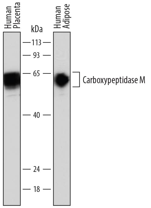
Detection of Human Carboxypeptidase M by Western Blot.
Western blot shows lysates of human placenta tissue and human adipose tissue. PVDF membrane was probed with 1 µg/mL of Sheep Anti-Human Carboxypeptidase M Antigen Affinity-purified Polyclonal Antibody (Catalog # AF7457) followed by HRP-conjugated Anti-Sheep IgG Secondary Antibody (Catalog # HAF016). A specific band was detected for Carboxypeptidase M at approximately 58-65 kDa (as indicated). This experiment was conducted under reducing conditions and using Immunoblot Buffer Group 1.Carboxypeptidase M in THP‑1 Human Cell Line.
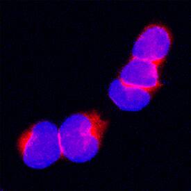
Carboxypeptidase M was detected in immersion fixed THP-1 human acute monocytic leukemia cell line using Sheep Anti-Human Carboxypeptidase M Antigen Affinity-purified Polyclonal Antibody (Catalog # AF7457) at 15 µg/mL for 3 hours at room temperature. Cells were stained using the NorthernLights™ 557-conjugated Anti-Sheep IgG Secondary Antibody (red; Catalog # NL010) and counterstained with DAPI (blue). Specific staining was localized to plasma membrane and cytoplasm. View our protocol for Fluorescent ICC Staining of Non-adherent Cells.Carboxypeptidase M in Human Placenta.
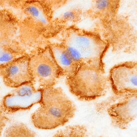
Carboxypeptidase M was detected in immersion fixed paraffin-embedded sections of human placenta using Sheep Anti-Human Carboxypeptidase M Antigen Affinity-purified Polyclonal Antibody (Catalog # AF7457) at 10 µg/mL overnight at 4 °C. Before incubation with the primary antibody, tissue was subjected to heat-induced epitope retrieval using Antigen Retrieval Reagent-Basic (Catalog # CTS013). Tissue was stained using the Anti-Sheep HRP-DAB Cell & Tissue Staining Kit (brown; Catalog # CTS019) and counterstained with hematoxylin (blue). Specific staining was localized to the plasma membrane of decidual cells. View our protocol for Chromogenic IHC Staining of Paraffin-embedded Tissue Sections.Applications for Human Carboxypeptidase M Antibody
Immunocytochemistry
Sample: Immersion fixed THP‑1 human acute monocytic leukemia cell line
Immunohistochemistry
Sample: Immersion fixed paraffin-embedded sections of human placenta subjected to heat-induced epitope retrieval using Antigen Retrieval Reagent-Basic (Catalog # CTS013)
Western Blot
Sample: Human placenta tissue and human adipose tissue
Formulation, Preparation, and Storage
Purification
Reconstitution
Formulation
Shipping
Stability & Storage
- 12 months from date of receipt, -20 to -70 °C as supplied.
- 1 month, 2 to 8 °C under sterile conditions after reconstitution.
- 6 months, -20 to -70 °C under sterile conditions after reconstitution.
Background: Carboxypeptidase M
CPM (Carboxypeptidase M) is a 50‑65 kDa monomer that belongs to the regulatory CPN/E subfamily, peptidase M14 family of enzymes. It is widely expressed, being found on macrophages, fibroblasts, endothelial cells, oligodendrocytes and Schwann cells, dendritic cells, osteoblasts and bronchial epithelium. CPM is a GPI‑linked glycoprotein that is best known as a peptidase that cleaves basic amino acids (aa) from the carboxyterminal of a number of peptides, including EGF and bradykinin. It is also known to bind to apparent substrates and undergo a conformational change that links it with the kinin B1 GPCR, initiating signal transduction. Mature human CPM is 406 aa in length (aa 18-423). It contains one large enzymatic region (aa 19-310) and two critical glutamic acid residues at Glu260 and Glu264. Like many GPI‑linked proteins, CPM undergoes solubilization and is reportedly found in urine and amniotic fluid. Over aa 18-423 (mature CPM), human CPM shares 86% aa sequence identity with mouse CPM.
Alternate Names
Gene Symbol
UniProt
Additional Carboxypeptidase M Products
Product Documents for Human Carboxypeptidase M Antibody
Product Specific Notices for Human Carboxypeptidase M Antibody
For research use only

