Fibroblast Activation Protein alpha/FAP Antibody (BLR150J)
Novus Biologicals, part of Bio-Techne | Catalog # NBP3-14728

Key Product Details
Species Reactivity
Applications
Label
Antibody Source
Concentration
Product Specifications
Immunogen
Clonality
Host
Description
Scientific Data Images for Fibroblast Activation Protein alpha/FAP Antibody (BLR150J)
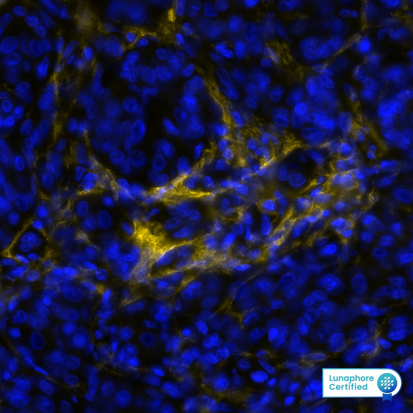
Detection of FAP in Human Melanoma via seqIF™ staining on COMET™
FAP was detected in immersion fixed paraffin-embedded sections of human Melanoma using Rabbit Anti-Human FAP, Monoclonal Antibody (Catalog# NBP3-14728) at 1:100 dilution at 37°Celsius for 4 minutes. Before incubation with the primary antibody, tissue underwent an all-in-one dewaxing and antigen retrieval preprocessing using PreTreatment Module (PT Module) and Dewax and HIER Buffer H (pH 9; Epredia Catalog # TA-999-DHBH). Tissue was stained using the Alexa Fluor™ Plus 647 Goat anti-Rabbit IgG Secondary Antibody at 1:200 at 37 ° Celsius for 2 minutes. (Yellow; Lunaphore Catalog # DR647RB) and counterstained with DAPI (blue; Lunaphore Catalog # DR100). Specific staining was localized to the membrane and cytoplasm of fibroblasts. Protocol available in COMET™ Panel Builder.Detection of FAP in Human Colon Cancer via seqIF™ staining on COMET™
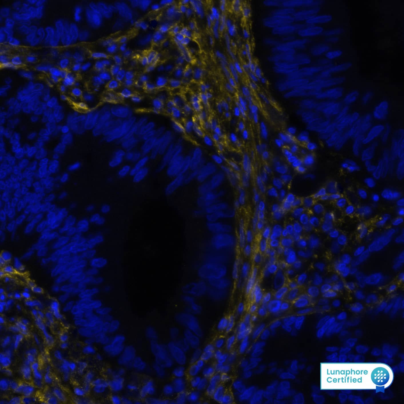
FAP was detected in immersion fixed paraffin-embedded sections of human Colon Cancer using Rabbit Anti-Human FAP, Monoclonal Antibody (Catalog# NBP3-14728) at 1:100 dilution at 37°Celsius for 4 minutes. Before incubation with the primary antibody, tissue underwent an all-in-one dewaxing and antigen retrieval preprocessing using PreTreatment Module (PT Module) and Dewax and HIER Buffer H (pH 9; Epredia Catalog # TA-999-DHBH). Tissue was stained using the Alexa Fluor™ Plus 647 Goat anti-Rabbit IgG Secondary Antibody at 1:200 at 37 ° Celsius for 2 minutes. (Yellow; Lunaphore Catalog # DR647RB) and counterstained with DAPI (blue; Lunaphore Catalog # DR100). Specific staining was localized to the membrane and cytoplasm of fibroblasts. Protocol available in COMET™ Panel Builder.Detection of FAP in Human Pancreas via seqIF™ staining on COMET™
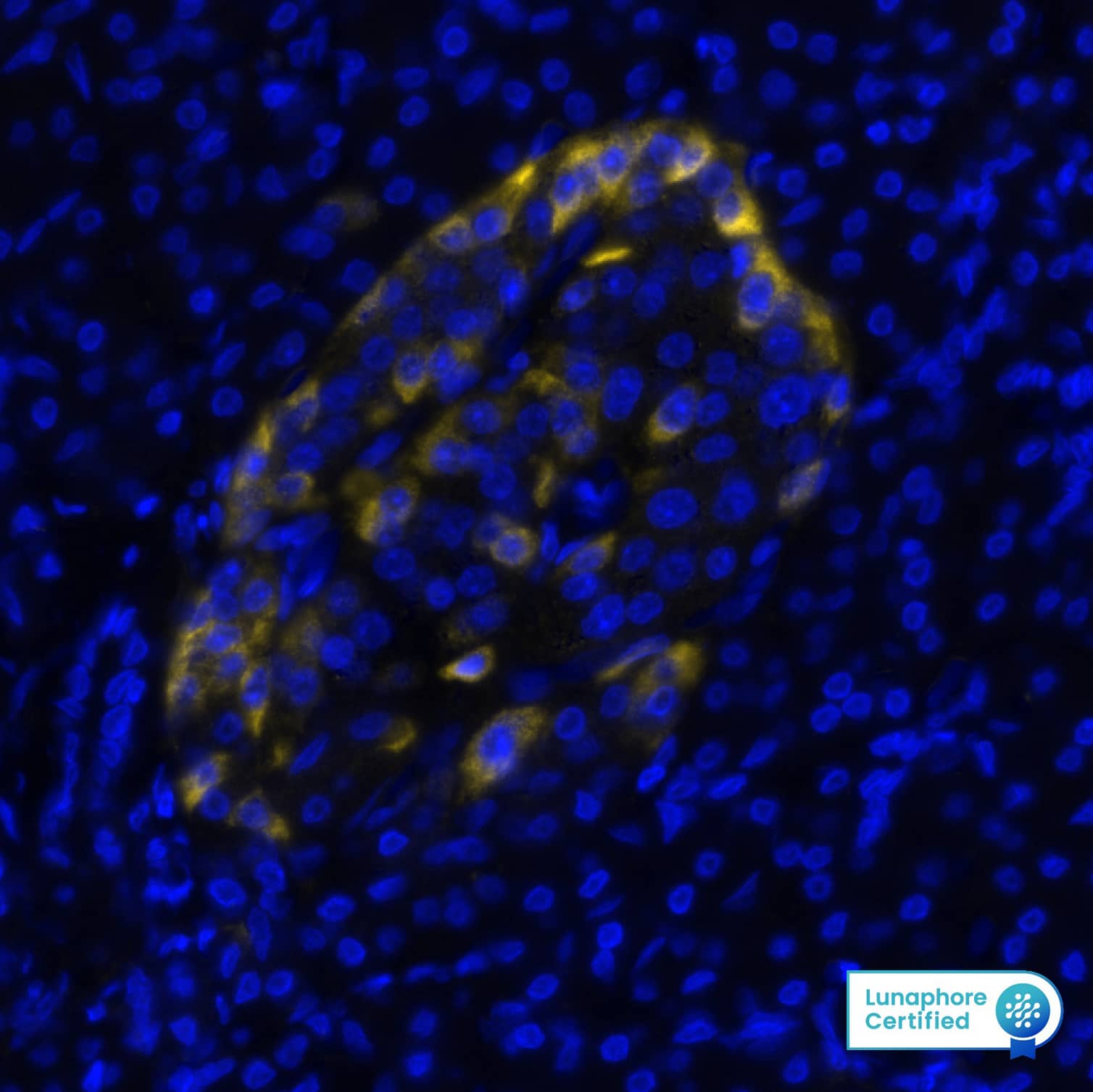
FAP was detected in immersion fixed paraffin-embedded sections of human Pancreas using Rabbit Anti-Human FAP, Monoclonal Antibody (Catalog# NBP3-14728) at 1:100 dilution at 37°Celsius for 4 minutes. Before incubation with the primary antibody, tissue underwent an all-in-one dewaxing and antigen retrieval preprocessing using PreTreatment Module (PT Module) and Dewax and HIER Buffer H (pH 9; Epredia Catalog # TA-999-DHBH). Tissue was stained using the Alexa Fluor™ Plus 647 Goat anti-Rabbit IgG Secondary Antibody at 1:200 at 37 ° Celsius for 2 minutes. (Yellow; Lunaphore Catalog # DR647RB) and counterstained with DAPI (blue; Lunaphore Catalog # DR100). Specific staining was localized to the cytoplasm of pancreatic islet cells. Protocol available in COMET™ Panel Builder.Applications for Fibroblast Activation Protein alpha/FAP Antibody (BLR150J)
Immunocytochemistry/ Immunofluorescence
Immunohistochemistry
Immunohistochemistry-Paraffin
Immunoprecipitation
Multiplex Immunofluorescence
Western Blot
Formulation, Preparation, and Storage
Purification
Formulation
Preservative
Concentration
Shipping
Stability & Storage
Background: Fibroblast Activation Protein alpha/FAP
FAP (also known as seprase) is a transmembrane serine protease, and a soluble and enzymatically active form of FAP known as antiplasmin-cleaving enzyme (APCE) circulates in human plasma. FAP is expressed on reactive stromal fibroblasts in tumor tissue and wound healing and on synoviocytes in rheumatoid arthritis. It exhibits dipeptidyl peptidase activity with substrate specificity similar to DPPIV/CD26, which is specific for N-terminal Xaa-Pro sequences. FAP is also an endopeptidase that can degrade Gelatin, Collagens I and IV, Fibronectin, and Laminin as well as several peptide hormones (e.g. Neuropeptide Y, Brain Natriuretic Peptide, Substance P, Peptide YY, and Incretins). The enzymatic activity is dependent on FAP association with DPPIV on the cell surface. The matrix-dedgrading activity of FAP contributes to tumor cell migration and invasion. In addition, FAP can enhance tumor cell growth by limiting the development of anti-tumor immunity.
Alternate Names
Gene Symbol
Additional Fibroblast Activation Protein alpha/FAP Products
- All Products for Fibroblast Activation Protein alpha/FAP
- Fibroblast Activation Protein alpha/FAP cDNA Clones
- Fibroblast Activation Protein alpha/FAP ELISA Kits
- Fibroblast Activation Protein alpha/FAP Lysates
- Fibroblast Activation Protein alpha/FAP Primary Antibodies
- Fibroblast Activation Protein alpha/FAP Proteins and Enzymes
- Fibroblast Activation Protein alpha/FAP Simple Plex
Product Documents for Fibroblast Activation Protein alpha/FAP Antibody (BLR150J)
Product Specific Notices for Fibroblast Activation Protein alpha/FAP Antibody (BLR150J)
This product is for research use only and is not approved for use in humans or in clinical diagnosis. Primary Antibodies are guaranteed for 1 year from date of receipt.


![Immunoprecipitation: Fibroblast Activation Protein alpha/FAP Antibody (BLR150J) [NBP3-14728] Immunoprecipitation: Fibroblast Activation Protein alpha/FAP Antibody (BLR150J) [NBP3-14728]](https://resources.bio-techne.com/images/products/Fibroblast-Activation-Protein-alpha-FAP-Antibody-Immunoprecipitation-NBP3-14728-img0001.jpg)

![Western Blot-Fibroblast Activation Protein alpha/FAP Antibody [NBP3-14728] - Fibroblast Activation Protein alpha/FAP Antibody (BLR150J)](https://resources.bio-techne.com/images/products/nbp3-14728_rabbit-fibroblast-activation-protein-alpha-fap-mab-western-blot-22320249348..jpg)
![Immunohistochemistry-Paraffin-Fibroblast Activation Protein alpha/FAP Antibody [NBP3-14728] - Fibroblast Activation Protein alpha/FAP Antibody (BLR150J)](https://resources.bio-techne.com/images/products/nbp3-14728_rabbit-fibroblast-activation-protein-alpha-fap-mab-immunohistochemistry-paraffin-22320249549..jpg)
![ICC/IF- Fibroblast Activation Protein alpha/FAP Antibody [NBP3-14728] - Fibroblast Activation Protein alpha/FAP Antibody (BLR150J)](https://resources.bio-techne.com/images/products/nbp3-14728_rabbit-fibroblast-activation-protein-alpha-fap-mab-immunocytochemistry-immunofluorescence-22320249755..jpg)