
COX-2 Antibody
Novus Biologicals, part of Bio-Techne | Catalog # NB100-868



Scientific images may be resized, cropped, or adjusted for brightness or contrast for presentation purposes. These updates are limited to presentation and do not affect the underlying scientific interpretation of the data. Where available, additional source data may be provided upon request.
Key Product Details
Species Reactivity
Validated:
Human, Mouse
Cited:
Human, Mouse, Plant
Applications
Validated:
Immunohistochemistry, Immunohistochemistry-Paraffin, Western Blot, Peptide ELISA, Flow Cytometry, Immunocytochemistry/ Immunofluorescence
Cited:
Immunohistochemistry-Paraffin, Western Blot
Label
Unconjugated
Antibody Source
Polyclonal Goat IgG
Concentration
0.5 mg/ml
Product Specifications
Immunogen
Peptide with sequence C-NPTVLLKERSTEL corresponding to C-Terminus according to NP_000954.1.
Reactivity Notes
Expected from sequence similarity: Canine, Porcine, Bovine. Mouse reactivity reported in scientific literature (PMID: 24854039).
Localization
Microsomal membrane
Clonality
Polyclonal
Host
Goat
Isotype
IgG
Scientific Data Images for COX-2 Antibody
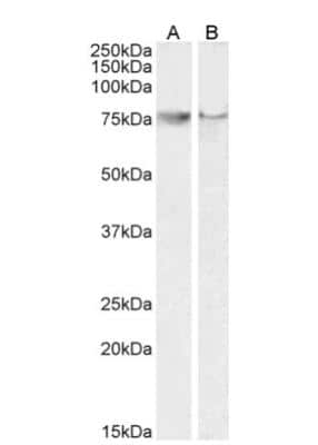
Western Blot: COX-2 Antibody [NB100-868]
Western Blot: COX-2 Antibody [NB100-868] - A549 (A) and Daudi (B) cell lysates (35 ug protein in RIPA buffer). Detected by chemiluminescence.
Immunocytochemistry/ Immunofluorescence: COX-2 Antibody [NB100-868]
Immunocytochemistry/Immunofluorescence: COX-2 Antibody [NB100-868] - Analysis of paraformaldehyde fixed NIH3T3 cells, permeabilized with 0.15%Triton. Primary incubation 1hr (10ug/ml) followed by Alexa Fluor 488 secondary antibody (2ug/ml), showing cytoplasm and vesicle staining. The nuclear stain is DAPI (blue). Negative control: Unimmunized goat IgG (10ug/ml) followed by Alexa Fluor 488 secondary antibody (2ug/ml).
Immunohistochemistry-Paraffin: COX-2 Antibody [NB100-868]
Immunohistochemistry-Paraffin: COX-2 Antibody [NB100-868] - 10x image of mouse lung tissue expressing cox-2. Secondary antibody: Alexa Fluor 546 (red), counterstain: DAPI (blue). Goat anti-mouse cox-2 immunofluorescence staining of paraffin embedded fixed mouse lung tissue sections. Antigen retrieval in citrate buffer (95C, 20 min) followed by permeabilization with 0.2% Triton X-100 for 20 min. Sections were blocked in 5% donkey serum for 1 hr at RT, and incubated with NB100-868 1:100 dilution in 3% donkey serum overnight at 4 C. Incubated with secondary donkey anti-goat AF546 at 1:500 dilution 1 hr at RT. Image submitted by a verified customer review.Applications for COX-2 Antibody
Application
Recommended Usage
Flow Cytometry
10ug/ml
Immunocytochemistry/ Immunofluorescence
10 ug/ml
Peptide ELISA
Detection limit 1:32000
Western Blot
0.1-0.3ug/ml
Application Notes
Western blot: Approx 75-80 kDa band observed in lysates of cell lines A549 and Daudi (predicted MW of 69 kDa according to NP_000954.1). COX-2 antibody validated for IHC-P from a verified customer review.
Reviewed Applications
Read 1 review rated 5 using NB100-868 in the following applications:
Formulation, Preparation, and Storage
Purification
Immunogen affinity purified
Formulation
Tris saline (20 mM Tris pH 7.3, 150 mM NaCl), 0.5% BSA
Preservative
0.02% Sodium Azide
Concentration
0.5 mg/ml
Shipping
The product is shipped with polar packs. Upon receipt, store it immediately at the temperature recommended below.
Stability & Storage
Store at -20C. Avoid freeze-thaw cycles.
Background: COX-2
Long Name
Cyclooxygenase 2
Alternate Names
COX2, PGHS-2, PHS-II, PTGS2
Entrez Gene IDs
5743 (Human)
Gene Symbol
PTGS2
UniProt
Additional COX-2 Products
Product Documents for COX-2 Antibody
Product Specific Notices for COX-2 Antibody
This product is for research use only and is not approved for use in humans or in clinical diagnosis. Primary Antibodies are guaranteed for 1 year from date of receipt.
Loading...
Loading...
Loading...
Loading...
Loading...
Loading...