
CD63 Antibody (H5C6) - BSA Free
Novus Biologicals, part of Bio-Techne | Catalog # NBP2-42225
Clone H5C6 was used by HLDA to establish CD designation.

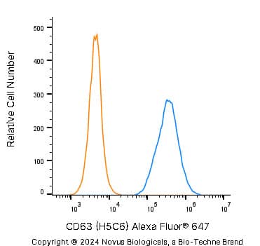
![Flow Cytometry: CD63 Antibody (H5C6) - BSA Free [NBP2-42225]](https://resources.bio-techne.com/images/products/CD63-Antibody-H5C6-Flow-Cytometry-NBP2-42225-img0013.jpg "Flow Cytometry: CD63 Antibody (H5C6) - BSA Free [NBP2-42225]")
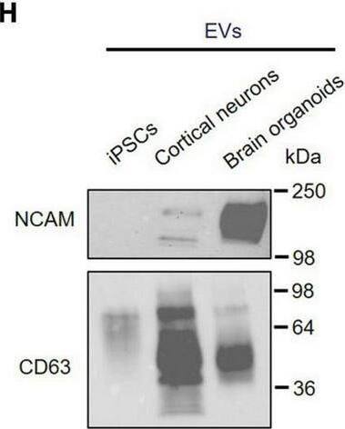
![Western Blot: CD63 Antibody (H5C6)BSA Free [NBP2-42225]](https://resources.bio-techne.com/images/products/CD63-Antibody-H5C6-Western-Blot-NBP2-42225-img0002.jpg "Western Blot: CD63 Antibody (H5C6)BSA Free [NBP2-42225]")
Scientific images may be resized, cropped, or adjusted for brightness or contrast for presentation purposes. These updates are limited to presentation and do not affect the underlying scientific interpretation of the data. Where available, additional source data may be provided upon request.
Conjugate
Catalog #
Forumulation
Catalog #
Key Product Details
Species Reactivity
Validated:
Human, Canine
Cited:
Human, Mouse, Rat, Canine
Applications
Validated:
Immunohistochemistry Whole-Mount, Western Blot, ELISA, Block/Neutralize, Flow Cytometry, Flow (Intracellular), Immunocytochemistry/ Immunofluorescence, Immunoprecipitation, Dot Blot, Functional, In vitro assay, Electron Microscopy
Cited:
Western Blot, ELISA, Immunoassay, Block/Neutralize, Flow Cytometry, Immunocytochemistry/ Immunofluorescence, Immunoprecipitation, Cytometric Bead Assay Standard, Dot Blot, In vitro assay, Functional Assay, Electron Microscopy
Label
Unconjugated
Antibody Source
Monoclonal Mouse IgG1 kappa Clone # H5C6
Format
BSA Free
Concentration
1.0 mg/ml
Product Specifications
Immunogen
Human splenic adherent cells.
Marker
Exosome Marker
Clonality
Monoclonal
Host
Mouse
Isotype
IgG1 kappa
Scientific Data Images for CD63 Antibody (H5C6) - BSA Free
Western Blot: CD63 Antibody (H5C6)BSA Free [NBP2-42225]
Western Blot: CD63 Antibody (H5C6) [NBP2-42225] - THP1 whole cell protein was separated by SDS-PAGE on a 12% gel and transferred to PVDF membrane. The membrane was probed with anti-CD63 antibody at 2 ug/mL and detected with an anti-mouse HRP secondary antibody using chemiluminescence.![Immunocytochemistry/ Immunofluorescence: CD63 Antibody (H5C6) - BSA Free [NBP2-42225]](https://resources.bio-techne.com/images/products/CD63-Antibody-H5C6-Immunocytochemistry-Immunofluorescence-NBP2-42225-img0001.jpg "Immunocytochemistry/ Immunofluorescence: CD63 Antibody (H5C6) - BSA Free [NBP2-42225]")
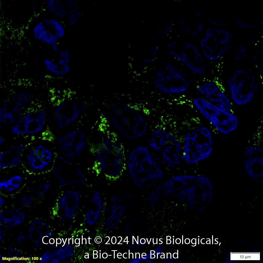
Immunocytochemistry/ Immunofluorescence: CD63 Antibody (H5C6) - BSA Free [NBP2-42225]
Immunocytochemistry/Immunofluorescence: CD63 Antibody (H5C6) [NBP2-42225] - The CD63 (H5C6) antibody was tested in HeLa cells at a 1:50 dilution against DyLight 488 (Green). Actin and nuclei were counterstained against Phalloidin 568 (Red) and DAPI (Blue), respectively.![Immunocytochemistry/ Immunofluorescence: CD63 Antibody (H5C6) - BSA Free [NBP2-42225]](https://resources.bio-techne.com/images/products/CD63-Antibody-H5C6-Immunocytochemistry-Immunofluorescence-NBP2-42225-img0014.jpg "Immunocytochemistry/ Immunofluorescence: CD63 Antibody (H5C6) - BSA Free [NBP2-42225]")
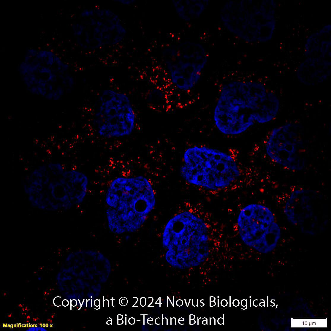
Immunocytochemistry/ Immunofluorescence: CD63 Antibody (H5C6) - BSA Free [NBP2-42225]
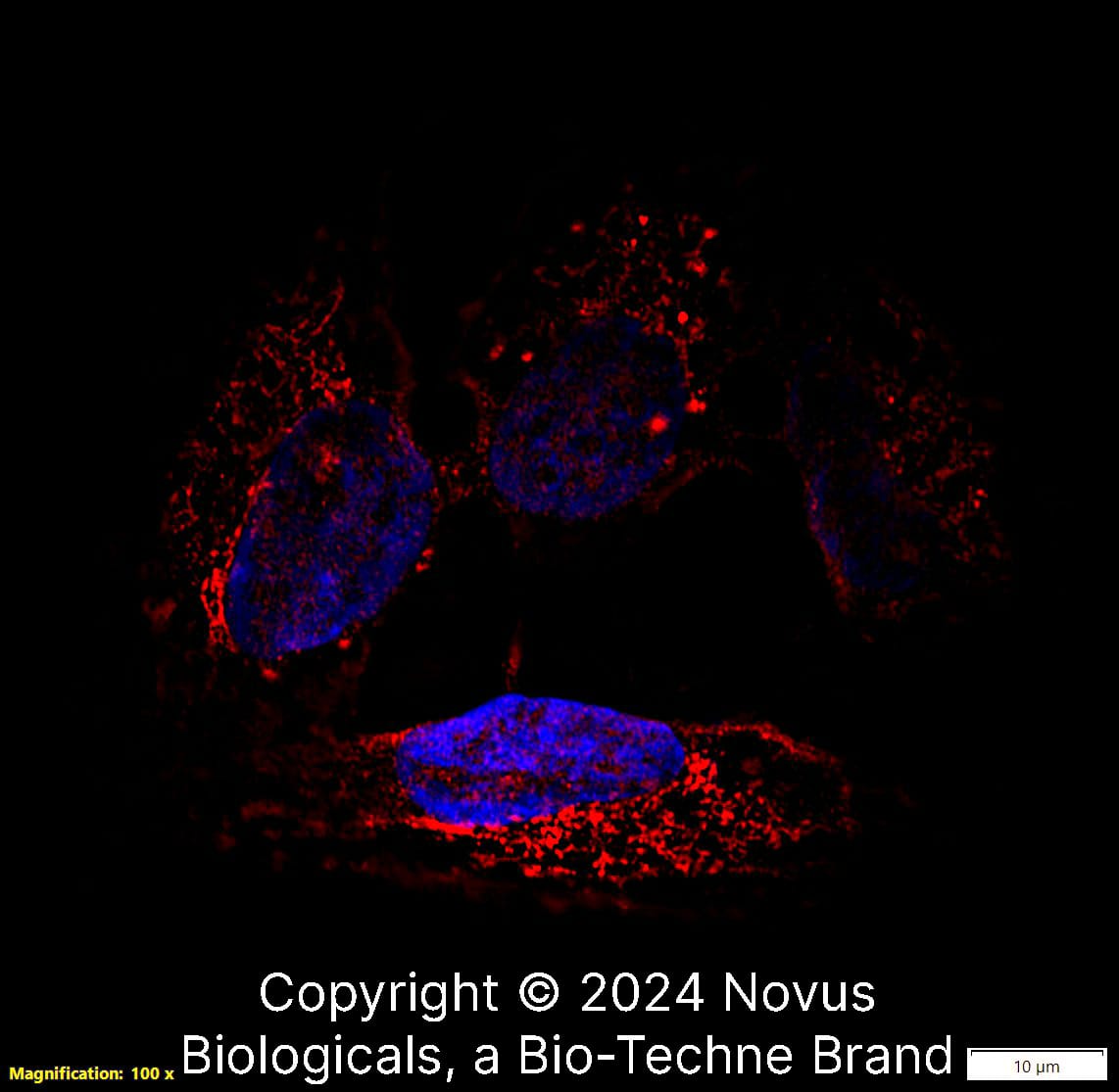
Immunocytochemistry/Immunofluorescence: CD63 Antibody (H5C6) [NBP2-42225] - HeLa cells were fixed in 4% paraformaldehyde for 10 minutes and permeabilized in 0.05% Triton X-100 in PBS for 5 minutes. The cells were incubated with anti-CD63 Antibody [H5C6] conjugated to DyLight 550 (NBP2-42225R) at 5 ug/ml for 1 hour at room temperature. Nuclei were counterstained with DAPI (Blue). Cells were imaged using a 100X objective and digitally deconvolved.![Immunocytochemistry/ Immunofluorescence: CD63 Antibody (H5C6) - BSA Free [NBP2-42225]](https://resources.bio-techne.com/images/products/CD63-Antibody-H5C6-Immunocytochemistry-Immunofluorescence-NBP2-42225-img0006.jpg "Immunocytochemistry/ Immunofluorescence: CD63 Antibody (H5C6) - BSA Free [NBP2-42225]")
![Immunocytochemistry/ Immunofluorescence: CD63 Antibody (H5C6) - BSA Free [NBP2-42225]](https://resources.bio-techne.com/images/products/CD63-Antibody-H5C6-Immunocytochemistry-Immunofluorescence-NBP2-42225-img0015.jpg "Immunocytochemistry/ Immunofluorescence: CD63 Antibody (H5C6) - BSA Free [NBP2-42225]")
![Immunocytochemistry/ Immunofluorescence: CD63 Antibody (H5C6) - BSA Free [NBP2-42225]](https://resources.bio-techne.com/images/products/CD63-Antibody-H5C6-Immunocytochemistry-Immunofluorescence-NBP2-42225-img0017.jpg "Immunocytochemistry/ Immunofluorescence: CD63 Antibody (H5C6) - BSA Free [NBP2-42225]")
![Immunocytochemistry/ Immunofluorescence: CD63 Antibody (H5C6) - BSA Free [NBP2-42225]](https://resources.bio-techne.com/images/products/CD63-Antibody-H5C6-Immunocytochemistry-Immunofluorescence-NBP2-42225-img0016.jpg "Immunocytochemistry/ Immunofluorescence: CD63 Antibody (H5C6) - BSA Free [NBP2-42225]")
![Immunocytochemistry/ Immunofluorescence: CD63 Antibody (H5C6) - BSA Free [NBP2-42225]](https://resources.bio-techne.com/images/products/CD63-Antibody-H5C6-Immunocytochemistry-Immunofluorescence-NBP2-42225-img0008.jpg "Immunocytochemistry/ Immunofluorescence: CD63 Antibody (H5C6) - BSA Free [NBP2-42225]")
![Western Blot: CD63 Antibody (H5C6)BSA Free [NBP2-42225]](https://resources.bio-techne.com/images/products/CD63-Antibody-H5C6-BSA-Free-Western-Blot-NBP2-42225-img0019.jpg "Western Blot: CD63 Antibody (H5C6)BSA Free [NBP2-42225]")
![Flow Cytometry: CD63 Antibody (H5C6) - BSA Free [NBP2-42225]](https://resources.bio-techne.com/images/products/CD63-Antibody-H5C6-Flow-Cytometry-NBP2-42225-img0018.jpg "Flow Cytometry: CD63 Antibody (H5C6) - BSA Free [NBP2-42225]")
![Western Blot: CD63 Antibody (H5C6)BSA Free [NBP2-42225]](https://resources.bio-techne.com/images/products/CD63-Antibody-H5C6-Western-Blot-NBP2-42225-img0007.jpg "Western Blot: CD63 Antibody (H5C6)BSA Free [NBP2-42225]")
![Immunocytochemistry/ Immunofluorescence: CD63 Antibody (H5C6) - BSA Free [NBP2-42225]](https://resources.bio-techne.com/images/products/CD63-Antibody-H5C6-Immunocytochemistry-Immunofluorescence-NBP2-42225-img0005.jpg "Immunocytochemistry/ Immunofluorescence: CD63 Antibody (H5C6) - BSA Free [NBP2-42225]")
![Flow Cytometry: CD63 Antibody (H5C6) - BSA Free [NBP2-42225]](https://resources.bio-techne.com/images/products/CD63-Antibody-H5C6-Flow-Cytometry-NBP2-42225-img0003.jpg "Flow Cytometry: CD63 Antibody (H5C6) - BSA Free [NBP2-42225]")
![Flow Cytometry: CD63 Antibody (H5C6) - BSA Free [NBP2-42225]](https://resources.bio-techne.com/images/products/CD63-Antibody-H5C6-Flow-Cytometry-NBP2-42225-img0004.jpg "Flow Cytometry: CD63 Antibody (H5C6) - BSA Free [NBP2-42225]")
![Flow (Intracellular): CD63 Antibody (H5C6) - BSA Free [NBP2-42225]](https://resources.bio-techne.com/images/products/CD63-Antibody-H5C6-Flow-Intracellular-NBP2-42225-img0009.jpg "Flow (Intracellular): CD63 Antibody (H5C6) - BSA Free [NBP2-42225]")
![Flow Cytometry: CD63 Antibody (H5C6) - BSA Free [NBP2-42225]](https://resources.bio-techne.com/images/products/CD63-Antibody-H5C6-Flow-Cytometry-NBP2-42225-img0010.jpg "Flow Cytometry: CD63 Antibody (H5C6) - BSA Free [NBP2-42225]")
![Flow Cytometry: CD63 Antibody (H5C6) - BSA Free [NBP2-42225]](https://resources.bio-techne.com/images/products/CD63-Antibody-H5C6-Flow-Cytometry-NBP2-42225-img0011.jpg "Flow Cytometry: CD63 Antibody (H5C6) - BSA Free [NBP2-42225]")
![Flow Cytometry: CD63 Antibody (H5C6) - BSA Free [NBP2-42225]](https://resources.bio-techne.com/images/products/CD63-Antibody-H5C6-Flow-Cytometry-NBP2-42225-img0012.jpg "Flow Cytometry: CD63 Antibody (H5C6) - BSA Free [NBP2-42225]")
Applications for CD63 Antibody (H5C6) - BSA Free
Application
Recommended Usage
Block/Neutralize
reported in scientific literature (PMID 35602933)
Electron Microscopy
reported in scientific literature (PMID 16735575)
Flow Cytometry
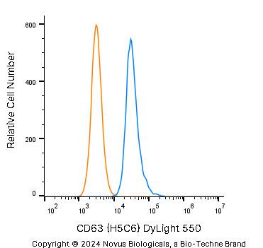
1:1000
Functional
reported in scientific literature (PMID 9811687)
Immunocytochemistry/ Immunofluorescence
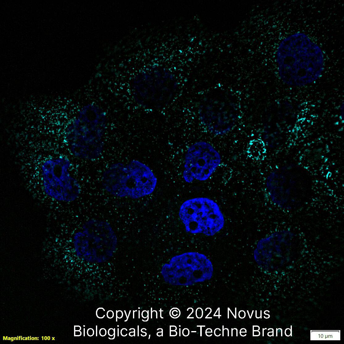
1:50-1:100
Immunohistochemistry Whole-Mount
reported in scientific literature (PMID 21464080)
In vitro assay
reported in scientific literature (PMID 21464080)
Reviewed Applications
Read 7 reviews rated 3.4 using NBP2-42225 in the following applications:
Formulation, Preparation, and Storage
Purification
Protein G purified
Formulation
PBS
Format
BSA Free
Preservative
0.05% Sodium Azide
Concentration
1.0 mg/ml
Shipping
The product is shipped with polar packs. Upon receipt, store it immediately at the temperature recommended below.
Stability & Storage
Store at 4C short term. Aliquot and store at -20C long term. Avoid freeze-thaw cycles.
Background: CD63
References
1. Pols, M. S., & Klumperman, J. (2009). Trafficking and function of the tetraspanin CD63. Experimental cell research. https://doi.org/10.1016/j.yexcr.2008.09.020
2. Metzelaar, M. J., Wijngaard, P. L., Peters, P. J., Sixma, J. J., Nieuwenhuis, H. K., & Clevers, H. C. (1991). CD63 antigen. A novel lysosomal membrane glycoprotein, cloned by a screening procedure for intracellular antigens in eukaryotic cells. The Journal of biological chemistry.
3. Horejsi, V., & Vlcek, C. (1991). Novel structurally distinct family of leucocyte surface glycoproteins including CD9, CD37, CD53 and CD63. FEBS letters. https://doi.org/10.1016/0014-5793(91)80988-f
4. Eckfeld, C., HauBler, D., Schoeps, B., Hermann, C. D., & Kruger, A. (2019). Functional disparities within the TIMP family in cancer: hints from molecular divergence. Cancer metastasis reviews. https://doi.org/10.1007/s10555-019-09812-6
5. Hoffmann, H. J., Santos, A. F., Mayorga, C., Nopp, A., Eberlein, B., Ferrer, M., Rouzaire, P., Ebo, D. G., Sabato, V., Sanz, M. L., Pecaric-Petkovic, T., Patil, S. U., Hausmann, O. V., Shreffler, W. G., Korosec, P., & Knol, E. F. (2015). The clinical utility of basophil activation testing in diagnosis and monitoring of allergic disease. Allergy. https://doi.org/10.1111/all.12698
6. Dell'Angelica, E. C., Shotelersuk, V., Aguilar, R. C., Gahl, W. A., & Bonifacino, J. S. (1999). Altered trafficking of lysosomal proteins in Hermansky-Pudlak syndrome due to mutations in the beta 3A subunit of the AP-3 adaptor. Molecular cell. https://doi.org/10.1016/s1097-2765(00)80170-7
Alternate Names
CD63, Granulophysin, Lamp-3, ME491, OMA81H, Tspan30
Entrez Gene IDs
967 (Human)
Gene Symbol
CD63
UniProt
Additional CD63 Products
Product Documents for CD63 Antibody (H5C6) - BSA Free
Product Specific Notices for CD63 Antibody (H5C6) - BSA Free
This product is for research use only and is not approved for use in humans or in clinical diagnosis. Primary Antibodies are guaranteed for 1 year from date of receipt.
Loading...
Loading...
Loading...
Loading...
Loading...