
CD43/Sialophorin Antibody (A2F8-R)
Novus Biologicals, part of Bio-Techne | Catalog # NBP3-32154
Recombinant Monoclonal Antibody

Key Product Details
Species Reactivity
Human
Applications
Immunohistochemistry, Immunohistochemistry-Paraffin, Western Blot, Immunocytochemistry/ Immunofluorescence
Label
Unconjugated
Antibody Source
Recombinant Monoclonal Mouse IgG1 Clone # A2F8-R expressed in HEK293
Concentration
1 mg/ml
Product Specifications
Immunogen
Synthetic peptide within C-terminal human CD43/Sialophorin. (Uniprot: P16150)
Localization
Membrane, microvillus, uropodium; Nucleus, PML body.
Clonality
Monoclonal
Host
Mouse
Isotype
IgG1
Theoretical MW
40 kDa.
Disclaimer note: The observed molecular weight of the protein may vary from the listed predicted molecular weight due to post translational modifications, post translation cleavages, relative charges, and other experimental factors.
Disclaimer note: The observed molecular weight of the protein may vary from the listed predicted molecular weight due to post translational modifications, post translation cleavages, relative charges, and other experimental factors.
Scientific Data Images for CD43/Sialophorin Antibody (A2F8-R)
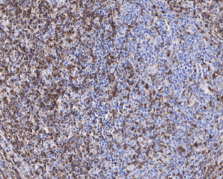
Immunohistochemistry: CD43/Sialophorin Antibody (A2F8-R) [NBP3-32154]
Immunohistochemistry: CD43/Sialophorin Antibody (A2F8-R) [NBP3-32154] - Immunohistochemical analysis of paraffin-embedded human B lymphocyte tumor tissue with Mouse anti-CD43/Sialophorin antibody (NBP3-32154) at 1/1,000 dilution.The section was pre-treated using heat mediated antigen retrieval with Tris-EDTA buffer (pH 9.0) for 20 minutes. The tissues were blocked in 1% BSA for 20 minutes at room temperature, washed with ddH2O and PBS, and then probed with the primary antibody (NBP3-32154) at 1/1,000 dilution for 1 hour at room temperature. The detection was performed using an HRP conjugated compact polymer system. DAB was used as the chromogen. Tissues were counterstained with hematoxylin and mounted with DPX.
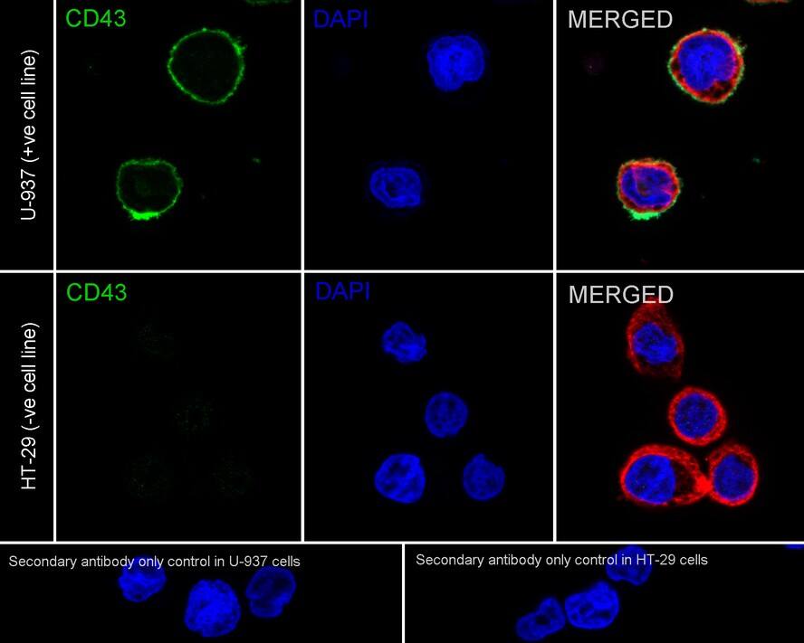
Immunocytochemistry/ Immunofluorescence: CD43/Sialophorin Antibody (A2F8-R) [NBP3-32154]
Immunocytochemistry/ Immunofluorescence: CD43/Sialophorin Antibody (A2F8-R) [NBP3-32154] - Immunocytochemistry analysis of U-937 (positive) and HT-29 (negative) labeling CD43/Sialophorin with Mouse anti-CD43/Sialophorin antibody (NBP3-32154) at 1/250 dilution.Cells were fixed in 4% paraformaldehyde for 20 minutes at room temperature, permeabilized with 0.1% Triton X-100 in PBS for 5 minutes at room temperature, then blocked with 1% BSA in 10% negative goat serum for 1 hour at room temperature. Cells were then incubated with Mouse anti-CD43/Sialophorin antibody (NBP3-32154) at 1/250 dilution in 1% BSA in PBST overnight at 4 ℃. Goat Anti-Mouse IgG H&L (iFluor™ 488) was used as the secondary antibody at 1/1,000 dilution. PBS instead of the primary antibody was used as the secondary antibody only control. Nuclear DNA was labelled in blue with DAPI.
beta Tubulin (red) was stained at 1/100 dilution overnight at +4℃. Goat Anti-Rabbit IgG H&L (iFluor™ 594) were used as the secondary antibody at 1/1,000 dilution.
![CD43/Sialophorin Antibody (A2F8-R) Western Blot: CD43/Sialophorin Antibody (A2F8-R) [NBP3-32154]](https://resources.bio-techne.com/images/products/nbp3-32154_-western-blot-639204590079360154.jpg "Western Blot: CD43/Sialophorin Antibody (A2F8-R) [NBP3-32154]")
Western Blot: CD43/Sialophorin Antibody (A2F8-R) [NBP3-32154]
Western blot analysis on different lysates with NBP3-32154 at 1/1,000 dilution. Lane 1: K-562 cell lysate Lane 2: HL-60 cell lysate Lane 3: THP-1 cell lysate Lane 4: U-937 cell lysate Lane 5: HT-29 cell lysate (negative) Lysates/proteins at 20 µg/Lane. Predicted band size: 40 kDa Observed band size: 100-130 kDa Exposure time: 3 minutes; 4-20% SDS-PAGE gel. Proteins were transferred to a PVDF membrane and blocked with 5% NFDM/TBST for 1 hour at room temperature. NBP3-32154 at 1/1,000 dilution was used in 5% NFDM/TBST at 4℃ overnight. Goat anti-Mouse IgG [HRP] at 1/50,000 dilution was used for 1 hour at room temperature.Applications for CD43/Sialophorin Antibody (A2F8-R)
Application
Recommended Usage
Immunocytochemistry/ Immunofluorescence
1:250
Immunohistochemistry-Paraffin
1:1000
Western Blot
1:1000
Formulation, Preparation, and Storage
Purification
Protein A purified
Formulation
PBS (pH7.4), 0.1% BSA and 40% Glycerol
Preservative
0.05% Sodium Azide
Concentration
1 mg/ml
Shipping
The product is shipped with polar packs. Upon receipt, store it immediately at the temperature recommended below.
Stability & Storage
Store at 4C short term. Aliquot and store at -20C long term. Avoid freeze-thaw cycles.
Background: CD43
Long Name
Cluster of Differentiation 43
Alternate Names
CD43, GPL115, Leukosialin, LSN, Ly-48, Sialophorin, SPN
Gene Symbol
SPN
Additional CD43 Products
Product Documents for CD43/Sialophorin Antibody (A2F8-R)
Product Specific Notices for CD43/Sialophorin Antibody (A2F8-R)
This product is for research use only and is not approved for use in humans or in clinical diagnosis. Primary Antibodies are guaranteed for 1 year from date of receipt.
Loading...
Loading...
Loading...
Loading...